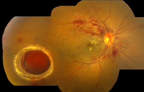
Ruptured retinal artery macroaneurysm
Article Sidebar
Main Article Content
Abstract
A 50-year-old man presented at Rajan Eye Care Hospital, Chennai, India, with blurring of vision and headaches of 2 months’ duration. There was so significant past systemic or ocular history. His best-corrected visual acuity in each eye was 6/9. Intraocular pressure, anterior segment examination, and pupil reaction were normal. Dilated fundus examination showed normal-appearing disc in each eye, with multiple flame-shaped hemorrhages and hard exudates in the macula in the form of an incomplete macular star. The right eye showed a round, reddish elevated lesion with surrounding hard exudates suggestive of intraretinal bleeding. The patient’s blood pressure was 210/120 mm Hg. The patient was diagnosed with bilateral, grade 3 hypertensive retinopathy, with a right eye ruptured retinal artery macroaneurysm (RAM). The lesion was observed due to the peripheral location, and the patient was referred for systemic medical evaluation and further management. RAM occurs in the bifurcation of retinal arterioles because of thinning and loss of elasticity following chronic hypertension. It is commonly seen within the first three orders of bifurcations and in temporal quadrants. In our patient, ruptured RAM was the presenting feature of undiagnosed hypertension and was seen in an uncommon location—the inferotemporal midperipheral retina.
Downloads
Article Details

This work is licensed under a Creative Commons Attribution-NonCommercial-NoDerivatives 4.0 International License.