Bilateral optic nerve head drusen
Article Sidebar
Main Article Content
Abstract
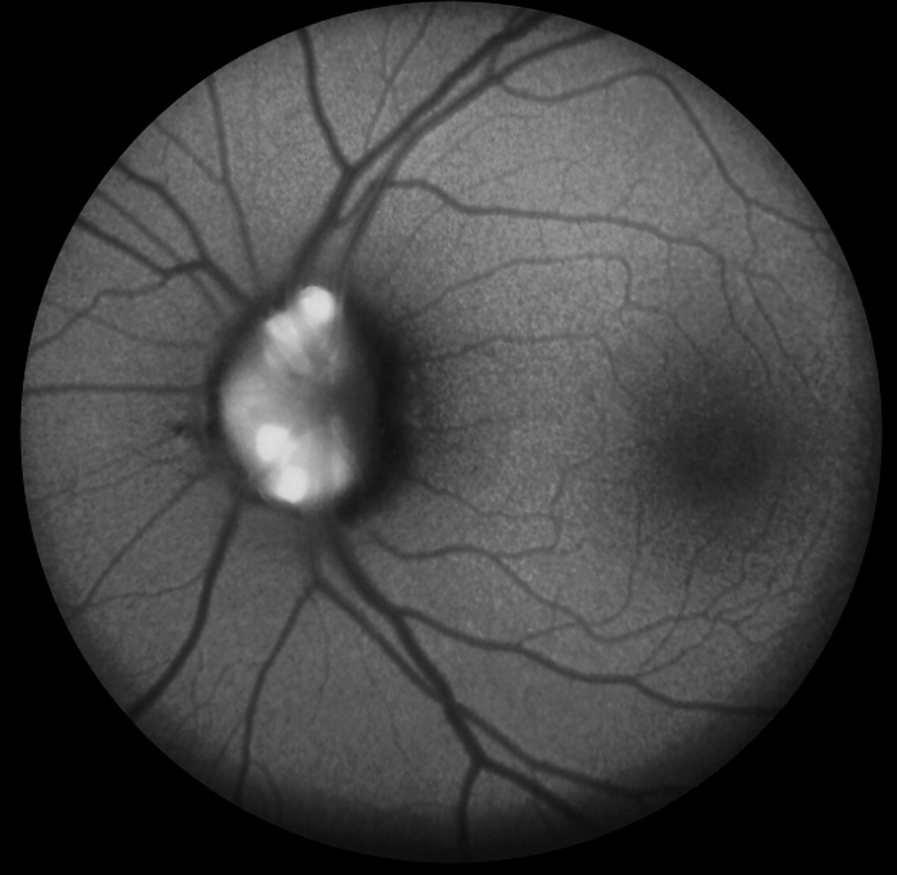
Fundus examination of a 27-year-old woman with best-corrected visual acuity of 6/6, N6 and normal intraocular pressure in each eye revealed small, crowded discs with a lumpy-bumpy appearance of the optic nerve (A-B). Visual fields performed elsewhere showed enlargement of the blind spot in the left eye. Fundus autofluorescence revealed hyperautofluorescent deposits suggestive of optic nerve head drusen (ONHD) in each eye, more in the left eye (C-D). ONHD and glaucoma can have similar visual field defects. The risk for visual field loss is higher with denser drusen and an obscured optic cup. Glaucomatous field defects in eyes without cupping should raise the suspicion of ONHD.
Downloads
Article Details

This work is licensed under a Creative Commons Attribution-NonCommercial-NoDerivatives 4.0 International License.