A simple eye model for practicing indirect ophthalmoscopy and retinal laser photocoagulation
Article Sidebar
Main Article Content
Abstract
Purpose
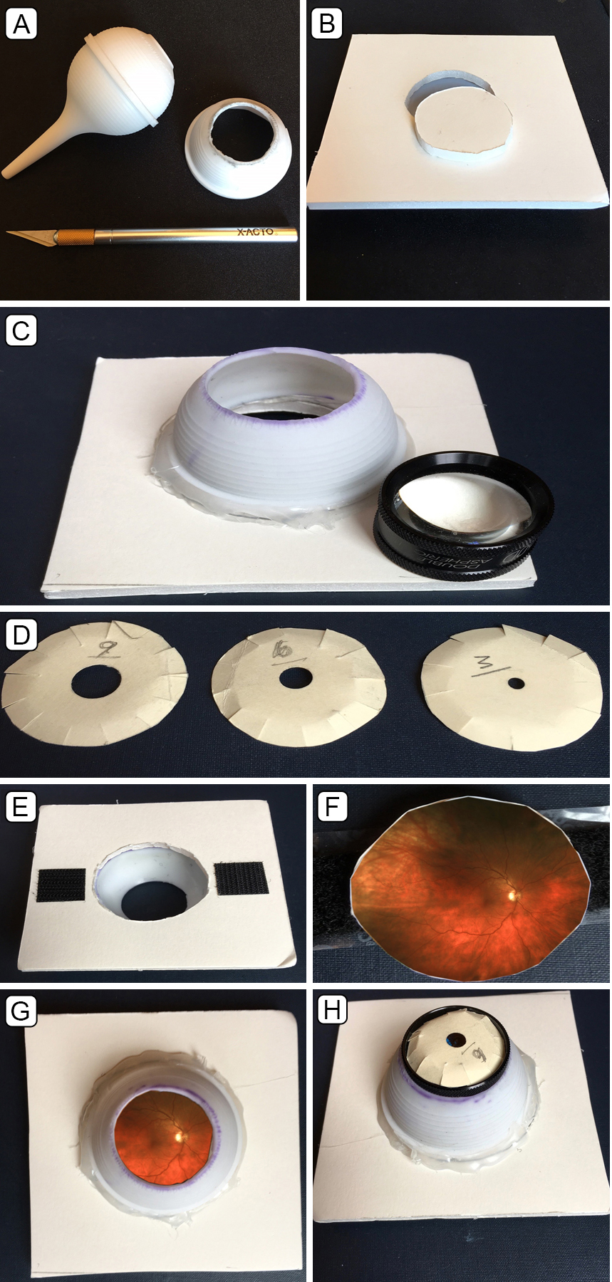
To describe a simple and inexpensive model eye that allows lifelike simulation of indirect ophthalmoscopy and retinal photocoagulation.
Methods
A 60 D examination lens, a bulb syringe, foam poster-board, a manila folder, a hobby knife, a fine pair of scissors, a glue gun, and a 2.5 cm square Optos color fundus photograph printed at 1200 dpi resolution on glossy photographic paper were used to create a model eye.
Results
This model produces a high-quality, inverted, and aerial image that closely simulates clinical indirect ophthalmoscopy. Pupil size and retinal pathology can be easily changed. Binocular indirect laser photocoagulation can also be simulated, because white laser burns will appear on the glossy inkjet photograph.
Conclusions
Binocular indirect ophthalmoscopy and indirect laser photocoagulation are technically challenging diagnostic and therapeutic techniques. This simple and easy-to-build eye model allows for lifelike simulation of indirect ophthalmoscopy and indirect laser retinal photocoagulation.
Downloads
Article Details

This work is licensed under a Creative Commons Attribution-NonCommercial-NoDerivatives 4.0 International License.


