Bilateral multifocal choroidal osteoma with choroidal neovascular membrane
Article Sidebar
Published:
Jan 13, 2019
Main Article Content
Abstract
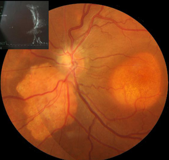
A healthy 32-year-old woman presented at Aravind Eye Hospital, Pondicherry, with gradual vision loss in her right eye (20/80) of 1 year’s duration. Fundus examination of the right eye showed two discrete yellowish-white subretinal lesions in the peripapillary area (A), pigmentary changes of the macula, and a dull foveal reflex. The fundus examination of the asymptomatic left eye showed a similar B-scan ultrasonography revealed hypereflectivity at the choroidal level that corresponded to the lesions, with back shadowing confirming the lesions’ calcific nature, suggestive of bilateral, multifocal choroidal osteoma (A-B, inset). Optical coherence tomography showed a choroidal neovascular membrane (CNVM), with intraretinal fluid in the right eye (C) and a dry macula in left eye (D). Investigations for secondary causes of the calcific deposits were negative, including calcium levels, thyroid profile, and blood counts. The patient received 2 doses of intravitreal bevacizumab. Following treatment, the CNVM remained inactive over 12 months’ follow-up (E and G). The left eye macula remained dry throughout the course of the disease (F and H).
Downloads
Download data is not yet available.
Article Details
How to Cite
1.
Krishnappa N, Ganne P. Bilateral multifocal choroidal osteoma with choroidal neovascular membrane. Digit J Ophthalmol. 2019;25(1). Accessed July 6, 2026. https://djo.harvard.edu/index.php/djo/article/view/251
Issue
Section
Images & Videos

This work is licensed under a Creative Commons Attribution-NonCommercial-NoDerivatives 4.0 International License.