Pseudo-Vossius ring after Descemet stripping automated endothelial keratoplasty
Article Sidebar
Main Article Content
Abstract
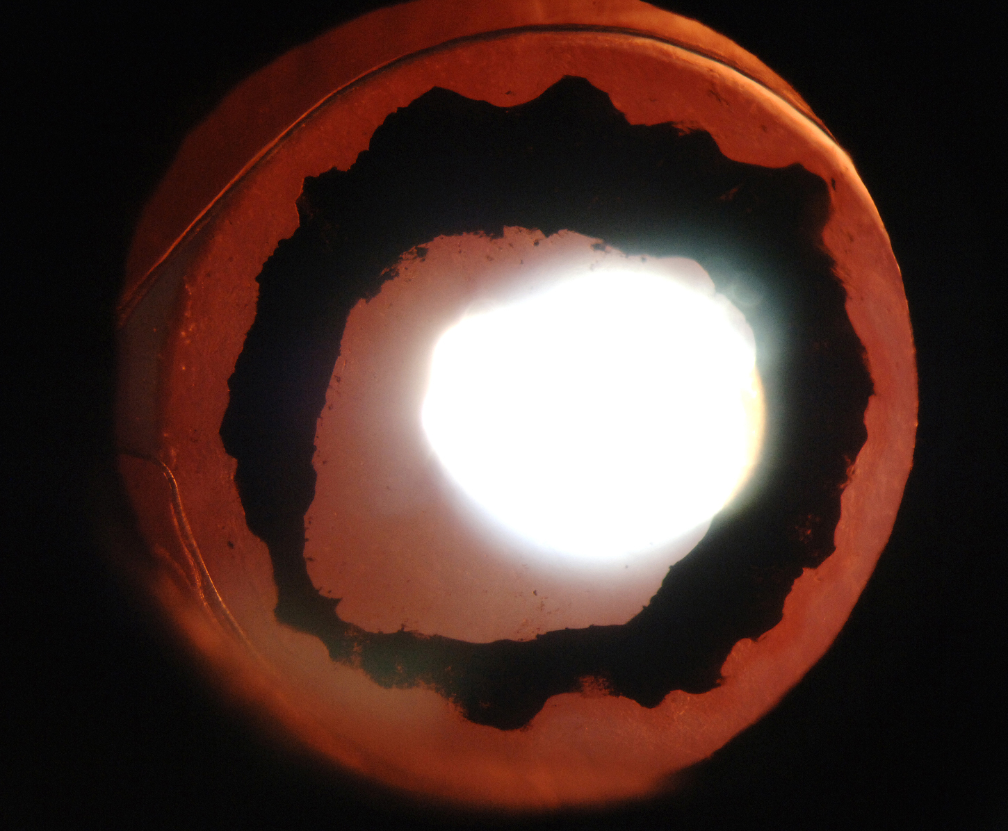
A 69-year-old, pseudophakic woman with Fuchs endothelial dystrophy and treated ocular hypertension underwent uncomplicated, planned Descemet stripping automated endothelial keratoplasty (DSAEK) at the Princess of Wales Hospital, Bridgend, 6 weeks after cataract extraction in the right eye. According to standard surgical protocol, after the endothelial graft was well centered, the anterior chamber was completely filled with air. After 5 minutes some of the air was released, leaving the anterior chamber filled with air to 60%. The patient was asked to remain supine for 24 hours postoperatively. One day after surgery, intraocular pressure measured 10 mm Hg in the operated eye. One week after surgery, dilated funduscopic examination showed a 360-degree, pigmented, pseudo-Vossius ring on the anterior lens capsule in the pseudophakic eye (A; retroillumination, B). There was no loss of iris pigmentation from the pupil edge; only the inferior peripheral iridotomy was transilluminating (C). The patient recovered without complication, with a best-corrected visual acuity of 6/9 (Snellen) at follow-up of 1 year. Pigment on the lens capsule persisted but did not cause visual symptoms. Vossius rings are usually noted on anterior lens capsule in phakic patients after blunt trauma. Pigmentation on the anterior lens capsule has also been noted to occur after endothelial grafts in phakic patients; our patient, however, was pseudophakic.
Downloads
Article Details

This work is licensed under a Creative Commons Attribution-NonCommercial-NoDerivatives 4.0 International License.