Proliferative diabetic retinopathy
Article Sidebar
Main Article Content
Abstract
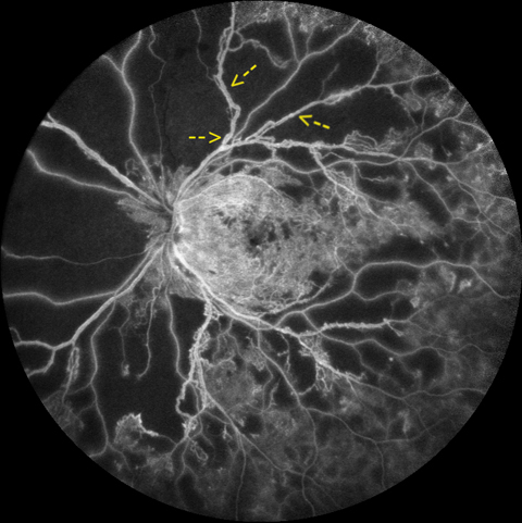
This fluorescein angiography image demonstrates advanced proliferative diabetic retinopathy (PDR) in a patient with poorly controlled type 1 diabetes mellitus. The image highlights extensive neovascularization originating from the optic disc, vessel infiltration of the macula, and diffuse neovascularization peripherally. Abundant neovascular nets, fluorescein leakage from these webs of thin, irregular, vessels are consistent with pathological angiogenesis. Additionally, 360° capillary nonperfusion is evident, contributing to retinal ischemia and the drive for neovascularization. The image also shows intraretinal microvascular abnormalities, vessel looping, and venous beading (yellow arrows). The pathophysiology of PDR involves chronic hyperglycemia-induced damage to retinal capillaries, leading to hypoxia-driven upregulation of vascular endothelial growth factor.
Downloads
Article Details

This work is licensed under a Creative Commons Attribution-NonCommercial-NoDerivatives 4.0 International License.