Vitreous cyst in a 73-year-old man
Article Sidebar
Main Article Content
Abstract
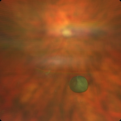
Intraocular cysts are rare. They may be classified into three subtypes, according to location: anterior chamber cysts, retrolental cysts (occurring between the lens and the hyaloid membrane), and vitreous cysts (VCs). VCs may be congenital, acquired, or idiopathic. Congenital VCs originate from epithelial elements, such as the ciliary body or remnants of the tunica vasculosa lentis. Acquired VCs may originate from retinal pathologies, such as uveitis, infections, retinoschisis, or retinitis pigmentosa. In most cases, VCs are an asymptomatic, incidental finding. In symptomatic patients, however, where VCs obscure the visual axis, photocystotomy or vitrectomy can be considered. Treatment depends on the severity of symptoms, because it can induce a series of iatrogenic complications, including cataracts. Slit-lamp photography, color fundus photography, B-scan ultrasonography, and/or ultrasound biomicroscopy are useful to monitor the position and, eventually, the dimension of the VCs. The image shows a solitary translucent, brown-pigmented VC that was observed in the anterior vitreous cavity in a 73-year-old man who presented, asymptomatic, to the University Eye Clinic of Trieste for routine ophthalmologic examination. In the absence of associated ocular or systemic conditions, the lesion was considered idiopathic, and we opted for conservative management with clinical monitoring.
Downloads
Article Details

This work is licensed under a Creative Commons Attribution-NonCommercial-NoDerivatives 4.0 International License.
References
N/A
Image