Subconjunctival cysticercosis
Article Sidebar
Main Article Content
Abstract
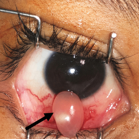
An 11-year-old boy presented at Advanced Eye Center, Post Graduate Institute of Medical Education and Research, Chandigarh, India, with a 1-month history of periocular swelling and pain in his right eye. Visual acuity was 6/6 in each eye. Examination revealed a 2 × 2 cm soft, cystic subconjunctival swelling in the right lower lid with injected overlying conjunctiva (A, arrows). Extraocular movements were full and unrestricted bilaterally. The remainder of the ocular examination was normal. T2-weighted magnetic resonance imaging (MRI) of the orbits showed a hyperintense lesion with a hypointense center, suggestive of cysticercosis. An MRI of the brain showed no evidence of neurocysticercosis. Excisional biopsy confirmed the presence of a cysticercosis cyst, and the periocular swelling resolved entirely after excision. Histopathological examination revealed the larval form of cysticercosis, with a fibrous wall and a double-layered eosinophilic membrane containing duct-like invaginations filled with neutrophil-rich inflammatory infiltrates. Cysticercosis is caused by the larval form of the tapeworm Taenia solium, typically contracted through the ingestion of eggs from contaminated food or water. The larvae migrate to various tissues, including the brain, eyes, and muscles. In tropical regions, orbital and subconjunctival spaces are common sites for these cysts. Treatment involves antihelminthic drugs such as albendazole with steroids, but surgical excision is preferred for subconjunctival cysts. Early intervention generally results in a favorable prognosis.
Downloads
Article Details

This work is licensed under a Creative Commons Attribution-NonCommercial-NoDerivatives 4.0 International License.