A chondromyxoid fibroma of the orbit with fundus autofluorescence in choroidal folds
Article Sidebar
Main Article Content
Abstract
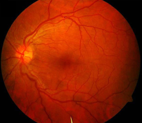
A 35-year-old woman presented at Advanced Eye Centre, PGIMER, Chandigarh, with left, painless proptosis and diminution of vision of 8 months duration. On examination, left inferior dystopia, proptosis of 4 mm on exophthalmometry, and gross upgaze restriction were noted. Palpation in the left superotemporal orbit revealed a firm, nontender orbital mass. Slit lamp fundus examination revealed multiple curvilinear lines that crossed the macula, superior to the left optic disc (A), and fundus autofluorescence revealed a hypo-hyper autofluorescent “zebra” pattern of lines highlighting the level beneath the retinal pigment epithelium choroid (B). A diagnosis of superior orbital mass with choroidal folds was made. Orbital computed tomography (C) revealed scleral indentation by the orbital mass (15 × 13 mm). Histopathology of the excised mass revealed a rare chondromyxoid fibroma of the orbit. The pathophysiology of chorioretinal folds includes wrinkling of Bruch’s membrane, retinal pigment epithelium, and adjacent choroid layers.
Downloads
Article Details

This work is licensed under a Creative Commons Attribution-NonCommercial-NoDerivatives 4.0 International License.