Straatsma syndrome: unilateral myelinated retinal nerve fibre layer, high myopia, and amblyopia
Article Sidebar
Main Article Content
Abstract
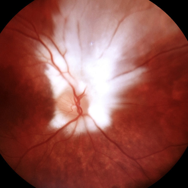
A 12-year-old boy was referred to the Ophthalmology Clinic at the State University of Campinas (UNICAMP) with a complaint of visual impairment in the right eye since childhood. On examination, the his best-corrected visual acuity was 20/30 in the right eye and 20/20 in the left eye. Cycloplegic refraction was −3.25 D in the right eye and −0.25 D in the left eye. Pupillary reflexes were equal and symmetric, with no relative afferent pupillary defect. Cover-uncover and alternate cover tests showed orthophoria for both distance and near fixation. Ocular motility was normal. Slit-lamp biomicroscopy examination was unremarkable. Dilated fundus examination revealed the presence of an extensive myelinated nerve fiber layer in the right eye surrounding the entire optic nerve, with feathery extensions superiorly (A). Dilated fundus examination of the left eye was normal (B). The patient was diagnosed with myelinated fibers, moderate unilateral myopia, and refractive amblyopia all of the right eye. Straatsma syndrome is a rare disease entity characterized by the classical triad of unilateral myelinated retinal nerve fibers, axial myopia, and amblyopia. Most patients have a poor visual prognosis. Therapeutic options include lenses for myopia correction and, when indicated, aggressive treatment for amblyopia.
Downloads
Article Details

This work is licensed under a Creative Commons Attribution-NonCommercial-NoDerivatives 4.0 International License.