40 year old woman with a five-day history of visual loss in the right eye
Article Sidebar
Main Article Content
Abstract
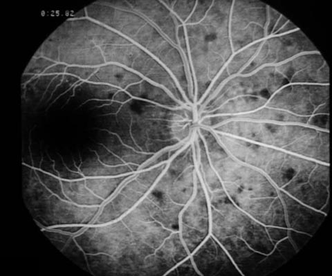
A 40-year-old white female presented to the Wills Eye Hospital Emergency Room with a five day history of blurred vision in the temporal visual field of her right eye. The patient denied any photopsia or other associated visual complaints.
Downloads
Article Details

This work is licensed under a Creative Commons Attribution-NonCommercial-NoDerivatives 4.0 International License.
References
Jampol LM, Sieving PA, Pugh D, Fishman GA, Gilbert H. Multiple evanescent white dot syndrome: I. Clinical findings. Arch Ophthalmol 1984;102:671-4.
Gass JDM. Acute posterior multifocal placoid pigment epitheliopathy. Arch Ophthalmol 1968;80:177-85.
Krill AE, Deutman AF. Acute retinal pigment epitheliitis. Am J Ophthalmol 1972;74:193-205.
Ryan SJ, Maumenee AE. Birdshot retinochoroidopathy. Am J Ophthalmol 1980;89:31-45.
Ie D, Glaser BM, Murphy RP, Gordon LW, Sjaarda RN, Thompson JT. Indocyanine green angiography in multiple evanescent white-dot syndrome. Am J Ophthalmol 1994;117:7-12.
Sieving PA, Fishman GA, Jampol LM, et al. Multiple evanescent white dot syndrome: II. Electrophysiology of the photoreceptots during retinal pigment epithelial disease. Arch Ophthalmol 1984;102:675-9.
Tsai L, Jampol LM, Pollock SC, et al. Chronic recurrent multiple evanescent white dot syndrome. Retina 1994;14:160-3.
Callanan D, Gass DM. Multifocal choroiditis and choroidal neovascularization associated with the multiple evanescent white dot and acute idiopathic blind spot enlargement syndrome. Ophthalmol 1992;99:1678-85.
Hamed LH, Glaser JS, Gass DM, et al. Protracted enlargement of the blind spot in multiple evanescent white dot syndrome. Arch Ophthalmol 1989;107:194-8.
Jampol LM: MEWDS, MFC, PIC, AMN, AIBSE, and AZOOR: one disease or many?. Retina 1995;15:373-8.