Dye-enhanced visualization of trabecular meshwork for canal-based minimally invasive glaucoma surgery
Main Article Content
Abstract
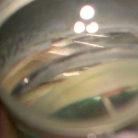
A male patient in his mid-fifties presented to our tertiary care hospital with diagnosis of both eyes moderate primary open angle glaucoma (POAG) with immature senile cataract (IMSC) was controlled on three antiglaucoma medications (AGM) in right eye (RE) and 2 AGMs in left eye (LE). On examination patient had best corrected visual acuity (BCVA) of 6/60 in RE with nuclear sclerosis of grade 2 along with a cup to disc ratio (CDR) of 0.8:1. LE examination showed a BCVA of 6/24 with nuclear sclerosis of grade 1 along with CDR of 0.8:1. On gonioscopy the angles were open in both the eyes up to scleral spur and the trabecular meshwork (TM) was lightly pigmented. Visual field examination by Humphrey Visual Field analyzer (Carl Zeiss Meditec,Dublin,CA,USA) showed moderate glaucoma in both eyes as per Hodapp -Parrish- Anderson criteria. Patient was planned for aqueous angiography guided BANG (Bent Ab-interno Needle Goniectomy) in high flow region followed by phacoemulsification surgery.[1] Indocyanine green (ICG) dye (0.5%) was injected in anterior chamber for performing aqueous angiography and simultaneous staining of the anterior capsule of the lens to facilitate capsulorrhexis.[1,2] On intraoperative gonioscopy it was noted that ICG dye also stained the TM to green color which made it easy to identify and incise the TM during BANG procedure (Figure 1A & 1B). We would like to highlight that ICG dye (0.5%) can aid in enhanced visualization of the TM and facilitate procedures of canal based MIGS by trainee surgeons (Figure 1B).
Downloads
Article Details

This work is licensed under a Creative Commons Attribution-NonCommercial-NoDerivatives 4.0 International License.
References
Dada T, Bukke AN. Aqueous angiography guided ab interno trabecular surgery for open-angle glaucoma. BMJ Case Rep. 2022 Jan 7;15(1):e248261.
Dada VK, Sharma N, Sudan R, Sethi H, Dada T, Pangtey MS. Anterior capsule staining for capsulorhexis in cases of white cataract: comparative clinical study. J Cataract Refract Surg. 2004 Feb;30(2):326-33.
