Atypical bilateral posterior polymorphous corneal dystrophy – a perpendicular presentation
Article Sidebar
Main Article Content
Abstract
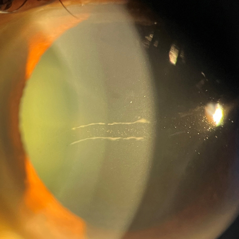
A 25-year-old woman presented at V N Desai Hospital, Mumbai, with occasional blurring of vision in both eyes. There was no history of forceps use or difficulty during normal delivery at birth. On examination, best-correct visual acuity in both eyes was 20/25. Slit-lamp examination of the right eye (A) showed two central, vertically oriented, parallel, bandlike lesions with serrated edges in the Descemet membrane. The left eye (B) had two central, horizontally oriented, parallel bands with a few fine vesicles in the intervening endothelial surface of the cornea. The remainder of the examination was unremarkable. The patient was diagnosed with posterior polymorphous corneal dystrophy (PPCD) in a variation where one eye has the classic horizontal orientation, and the other eye has a variant oriented vertically. PPCD is a rare, autosomal dominant corneal endothelial disorder with variable manifestations. Abnormal thickened Descemet membrane laid down by endothelial cells manifests as vesicular, bandlike or diffuse patterns at the posterior cornea. The exact incidence of PPCD is unknown because of its typically asymptomatic nature. Most patients have a bilateral, nonprogressive, asymptomatic disease, but asymmetric presentation is known to occur in many cases.
Downloads
Article Details

This work is licensed under a Creative Commons Attribution-NonCommercial-NoDerivatives 4.0 International License.