Laser-induced subhyaloidal and macular hemorrhage with complete recovery following vitrectomy
Article Sidebar
Main Article Content
Abstract
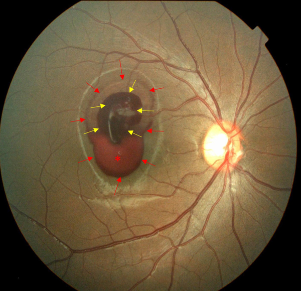
An 11-year-old boy presented with decreased vision after accidental exposure to a handheld blue laser pointer directed at his right eye. Best-corrected visual acuity in that eye was hand motions. Posterior segment examination revealed hemorrhaging in both the subhyaloidal space and the intramacular layers of the affected eye with sedimented blood in the subhyaloidal space. Raster OCT of the affected eye just above the sedimented blood level revealed two distinct membranes: a highly reflective membrane consistent with the ILM, and an overlying irregular, low-reflective membrane corresponding to the posterior hyaloid interface. A hyperreflective and well-defined lesion (clotted hemorrhage) was surrounded by hyperreflective dots (red blood cells) beyond the ILM. Additionally, a dome-shaped hyperreflective lesion corresponding to subhyaloidal hemorrhage was observed. Four days after the incident, the patient underwent a standard, 23-gauge, three-port, pars plana vitrectomy, with concurrent ILM peeling and aspiration of liquefied blood. Postoperatively, he was started on betamethasone eye drops 4 times daily for 2 weeks, followed by a taper. At 5 weeks’ follow-up, best-corrected visual acuity in the affected eye improved to 20/25, and fundus examination revealed complete resolution of premacular hemorrhage, without postoperative complications. Opacification of the RNFL was visible in the prefoveal area, but without obvious scarring. Corresponding OCT showed attenuation and irregularity of the inner retinal layers, including the RNFL, primarily on the temporal side of the fovea. The most likely source of hemorrhage in this case was a sub-branch of a retinal vessel, given the absence of any obvious chorioretinal laser burn damage.
Downloads
Article Details

This work is licensed under a Creative Commons Attribution-NonCommercial-NoDerivatives 4.0 International License.