The Versatile Teaching Eye: an affordable, 3D-printed model eye for simulating ophthalmic examination
Article Sidebar
Main Article Content
Abstract
Purpose
To describe the Versatile Teaching Eye (VT Eye), a 3D-printed model eye designed to provide an affordable examination simulator, and to report the results of a pilot program introducing the VT Eye and an ophthalmic training curriculum at a teaching hospital in Ghana.
Methods
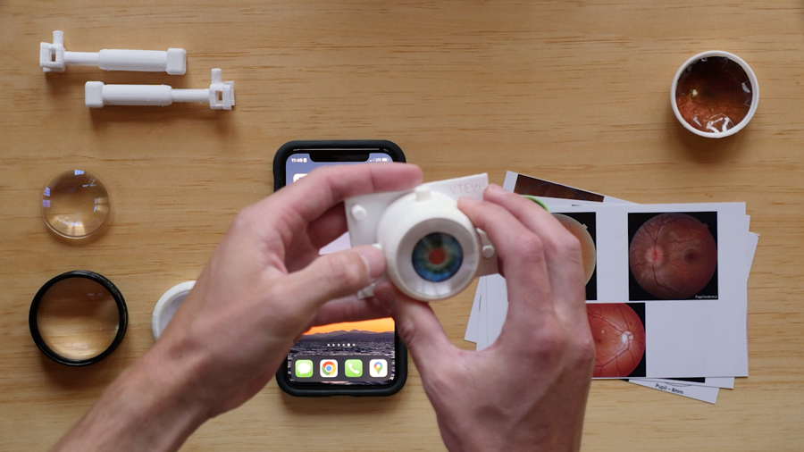
TinkerCAD was used to design the VT Eye, which was printed with ABS plastic. The design features an adapter that permits use of a smartphone as a digital fundus. We developed a set of digital flashcards allowing for an interactive review of a range of retinal pathologies. An analog fundus was developed for practicing traditional slit lamp and indirect examinations as well as retinal laser practice. The model was used for a period of 2 weeks by ophthalmic trainees at Komfo Anokye Teaching Hospital, Kumasi, Ghana, to practice indirect ophthalmoscopy, slit lamp biomicroscopy, smartphone funduscopy, and retinal image drawing. Results were assessed at by means of a pre-/post-training survey of 6 residents.
Results
The VT Eye accommodates diverse fundus examination techniques. Its 3D-printed design ensures cost-effective, high-quality replication. When paired with a 20 D practice examination lens, the digital fundus provides a comprehensive, interactive training environment for <$30.00 (USD). This device allows for indirect examination practice without requiring an indirect headset, which may increase the amount of available practice for trainees early in their careers. In the Ghana pilot program, the model’s use in indirect examination training sessions significantly boosted residents’ confidence in various examination techniques. Comparing pre- and post-session ratings, average reported confidence levels rose by 30% for acquiring clear views of the posterior pole, 42% for visualizing the periphery, and 141% for capturing important pathology using personal smartphones combined with a 20 D lens (all P < 0.05).
Conclusions
The VT Eye is readily reproducible and can be easily integrated into ophthalmic training curricula, even in regions with limited resources. It offers an effective and affordable training solution, underscoring its potential for global adoption and the benefits of incorporating innovative technologies in medical education.
Downloads
Article Details

This work is licensed under a Creative Commons Attribution-NonCommercial-NoDerivatives 4.0 International License.


References
Troncoso MU. New model of schematic eye for skiascopy (retinoscopy) and ophthalmoscopy. Am J Ophthalmol 1922;5:436-41. DOI: https://doi.org/10.1016/S0002-9394(22)90400-3
Chou J, Kosowsky T, Payal AR, Gonzalez Gonzalez LA, Daly MK. Construct and face validity of the Eyesi Indirect Ophthalmoscope Simulator. Retina 2017;37:1967. DOI: https://doi.org/10.1097/IAE.0000000000001438
Lee R, Raison N, Lau WY, et al. A systematic review of simulation-based training tools for technical and non-technical skills in ophthalmology. Eye 2020;34:1737-59. DOI: https://doi.org/10.1038/s41433-020-0832-1
Javaid M, Haleem A, Singh RP, Suman R. 3D printing applications for healthcare research and development. Global Health J 2022;6:217-26. DOI: https://doi.org/10.1016/j.glohj.2022.11.001
Lantz PE, Adams GGW. Postmortem monocular indirect ophthalmoscopy. J Forensic Sci 2005;50:1450-52. DOI: https://doi.org/10.1520/JFS2005137
Iqbal U. Smartphone fundus photography: a narrative review. Int J Retina Vitreous 2021;7:44. DOI: https://doi.org/10.1186/s40942-021-00313-9
Kylstra JA, Diaz JD. A simple eye model for practicing indirect ophthalmoscopy and retinal laser photocoagulation. Digit J Ophthalmol 2019;25:1-4. DOI: https://doi.org/10.5693/djo.01.2018.11.001
Lewallen S. A simple model for teaching indirect ophthalmoscopy. Br J Ophthalmol 2006;90:1328-9. DOI: https://doi.org/10.1136/bjo.2006.096784