Lipid keratopathy: clinicopathologic correlation
Article Sidebar

Main Article Content
Abstract
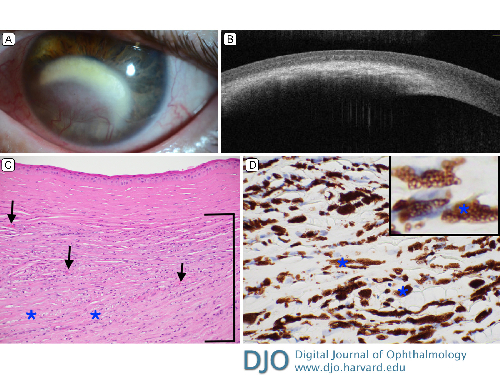 A 50-year-old woman presented at Massachusetts Eye and Ear with vision changes in her left eye of several months’ duration. Her medical history was significant for optic neuropathy in both eyes in the setting of chemotherapy and facial radiation for an unresectable sinonasal tumor. On examination, there was dense central, sea fan–shaped lipid keratopathy (A) of unknown etiology, although herpetic or post-radiation damage was in the differential diagnosis. Visual acuity in the left eye was hand motions. Optical coherence tomography revealed focal hyperreflectivity in the mid- and deep corneal stroma, corresponding to the lipid deposits seen clinically, with sparing of the epithelium and anterior stroma (B). She failed a trial of oral acyclovir and topical prednisolone, and penetrating keratoplasty was performed. Hematoxylin and eosin staining highlighted focal, pronounced mid- and deep stromal neovascularization, scarring, and foamy cells; the anterior stroma was spared (C; arrows mark blood vessels, blue asterisks mark foamy cells, and brackets outline the stromal scarring below the normal anterior stroma). Immunostaining for adipophilin accentuated the numerous foamy cells, highlighting lipid-containing vesicles (D; ×60 and inset). At 1 month postoperatively, her visual acuity was 20/200, with pinhole to 20/80.
Downloads
Article Details

This work is licensed under a Creative Commons Attribution-NonCommercial-NoDerivatives 4.0 International License.