Congenital simple hamartoma of the retinal pigment epithelium in an 11-year-old patient
Article Sidebar
Main Article Content
Abstract
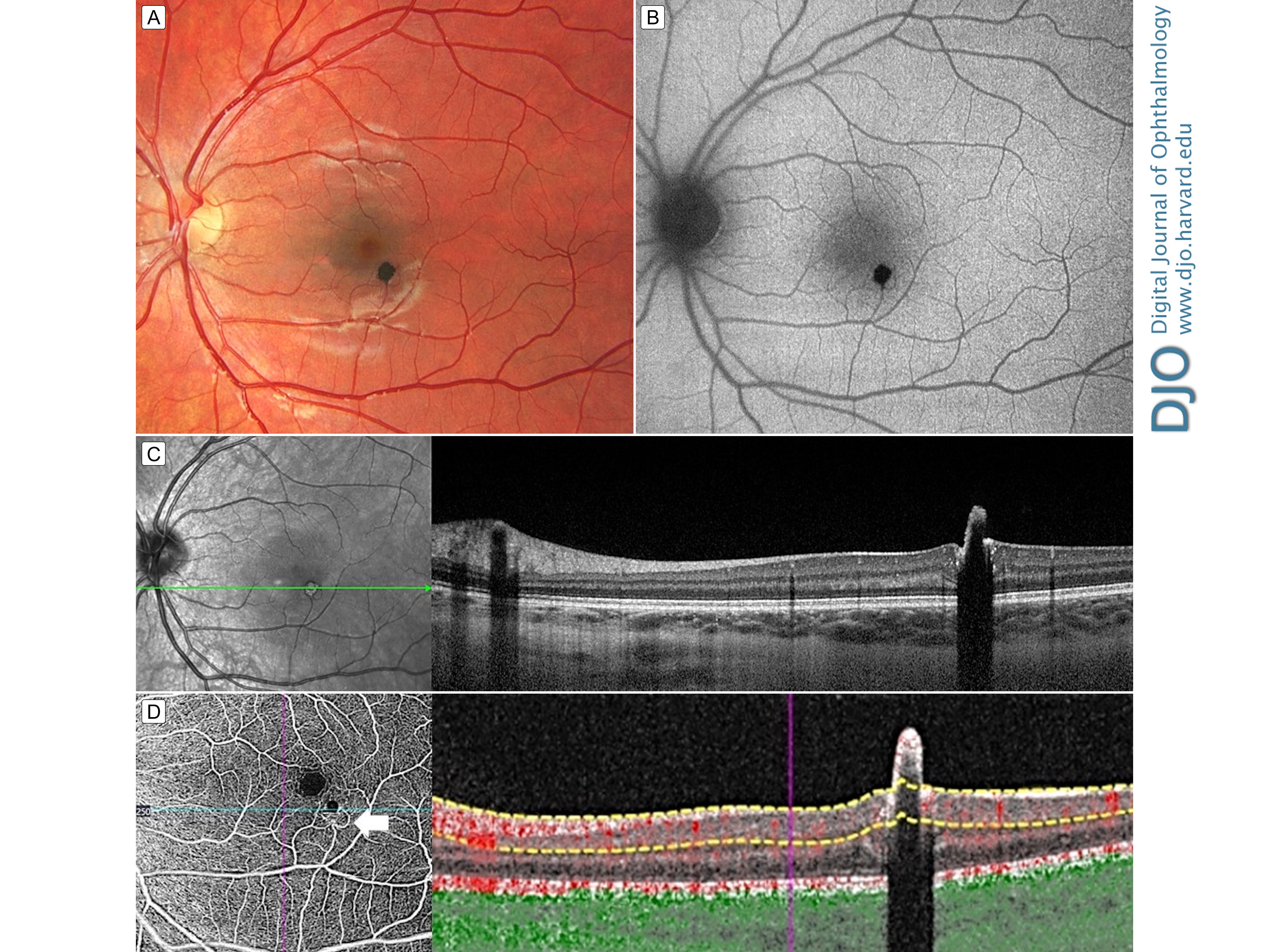
An asymptomatic 11-year-old girl presented at the Ocular Oncology Service, Wills Eye Hospital, for evaluation of a pigmented macular lesion in the left eye. On examination, visual acuity was 20/20 in each eye. Fundoscopy revealed a simple retinal pigment epithelium (RPE) hamartoma 300 µm inferior and temporal to the foveola and measuring 300 µm in diameter and 800 µm in thickness (A). The remaining retina was flat, with no edema. The hamartoma was hypoautofluorescent (B). Optical coherence tomography (OCT) revealed abrupt retinal elevation, with complete posterior shadowing (C), and OCT angiography showed vascular remodeling around the hamartoma. A feeding arteriole and draining venule emanated from the lesion (D, arrows). Congenital RPE hamartoma almost always manifests in the macula, rarely affects visual acuity if not located directly in the foveola, and has not been reported to enlarge or undergo malignant transformation. Support provided in part by the Eye Tumor Research Foundation, Philadelphia, PA (CLS).
Downloads
Article Details

This work is licensed under a Creative Commons Attribution-NonCommercial-NoDerivatives 4.0 International License.