Sea fan neovascularization in retinal detachment
Article Sidebar
Main Article Content
Abstract
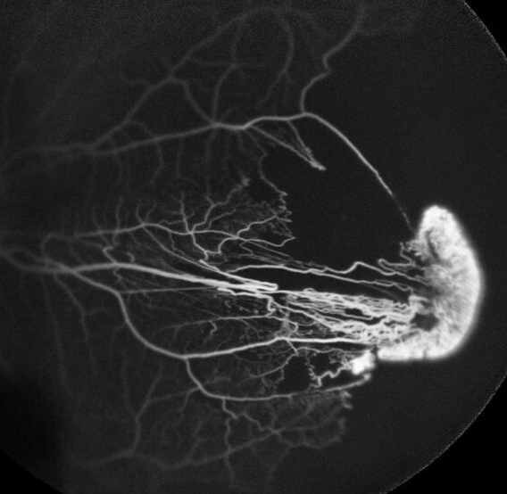
A 40-year-old healthy man presented at Sankara Nethralaya, Chennai, with sudden diminution of vision in the left eye of 10 days’ duration. On examination, best-corrected visual acuity in the left eye was 2/60 (N36). Slit-lamp biomicroscopy examination showed normal anatomy of the anterior segment. Dilated fundus examination revealed a temporal retinal detachment, with large peripheral neovascularization causing traction with sieve-like retinal holes posterior to neovascularization in the equatorial area (A). Fluorescein angiography revealed a sea fan–shaped neovascularization with leakage and peripheral capillary nonperfusion areas (B). After vitrectomy with silicone oil tamponade, his visual acuity improved to 6/36 (N18) with attached retina.
Downloads
Article Details

This work is licensed under a Creative Commons Attribution-NonCommercial-NoDerivatives 4.0 International License.