Persistent fetal vasculature on RetCam
Article Sidebar

Main Article Content
Abstract
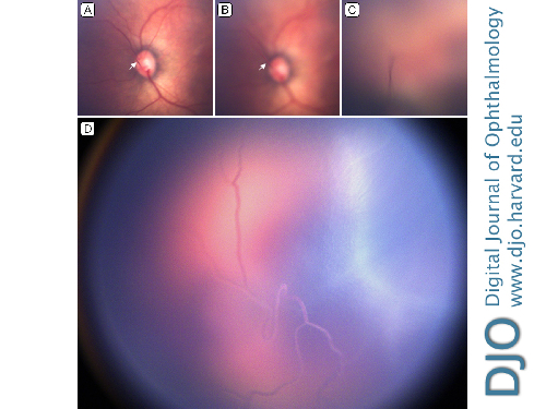
A premature neonate, born at 27 weeks’ gestational age and weighing 900 g, was referred for retinopathy of prematurity screening at Gold Coast University Hospital. The child was delivered by emergency cesarean section for fetal distress in the setting of maternal pyrexia. On RetCam imaging (Natus Medical Inc, Pleasanton, CA) at 36 weeks, a persistent hyaloid artery was seen extending from the optic nerve to the posterior lens surface, indicating persistent fetal vasculature (A-C). The branching vessels from the hyaloid artery were seen extending over the posterior surface of the lens capsule, with evidence of intravascular blood flow (D). There was no evidence of cataract, microphthalmia, or retinopathy of prematurity. Ongoing follow-up was arranged to monitor for cataract development.
Downloads
Article Details

This work is licensed under a Creative Commons Attribution-NonCommercial-NoDerivatives 4.0 International License.