Unilateral West Nile virus chorioretinitis in a 69-year-old woman
Article Sidebar
Main Article Content
Abstract
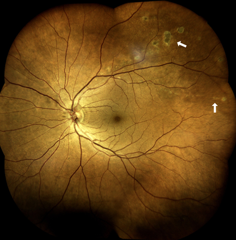
A 69-year-old woman was referred to our practice for investigation of new, circular, creamy chorioretinal lesions in a curvilinear distribution along the superotemporal vascular arcade of the left eye (A, arrows), found on routine eye examination. She endorsed a 1-week history of fevers and chills that self-resolved 1 month earlier. Fluorescein angiography of the left eye showed classic “target-like” staining of the lesions, characterized by a hypofluorescent center surrounded by a hyperfluorescent rim (B, arrows), prompting further history and workup for West Nile virus (WNV) chorioretinitis. History revealed daily mosquito bites, and serology for WNV returned positive IgM and IgG antibodies. There were no abnormal neurological findings. The patient was advised to avoid mosquitoes and subsequent follow-up over the next year showed no progression of the chorioretinal lesions. Chorioretinitis is a commonly overlooked presentation of WNV infection. Like most systemic manifestations of WNV, chorioretinitis tends to be self-limiting.
Downloads
Article Details

This work is licensed under a Creative Commons Attribution-NonCommercial-NoDerivatives 4.0 International License.