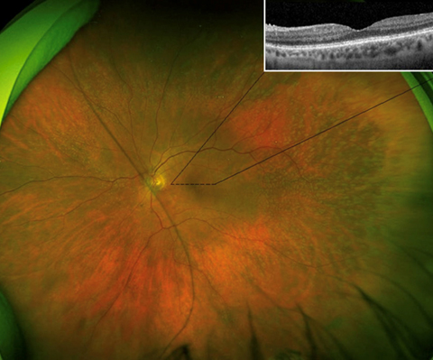
Widefield fundus photograph and optical coherence tomography of vitamin A deficiency retinopathy
Main Article Content
Abstract
A 55-year-old woman with end-stage nonalcoholic liver disease presented with nyctalopia. Her best-corrected visual acuity was 20/30 in both eyes. Funduscopic examination showed multiple, midperipheral yellow punctate deposits bilaterally. Optical coherence tomography of the macula revealed an attenuated ellipsoid zone in each eye (Figure, inset). Her vitamin A level was <5 mcg/dL (reference range, 32.5–78.0 mcg/dL). She started vitamin A and was advised to follow up in 4 weeks. Unfortunately, the patient was unable to follow up for repeat fundus examination. In cirrhosis, there is inadequate delivery of bile salts into the intestinal lumen, leading to insufficient absorption of fat-soluble vitamins such as vitamin A. Vitamin A, which is all-trans retinol, is a precursor of rhodopsin and a vital component of visual phototransduction. Thus, vitamin A deficiency commonly leads to nyctalopia. Supplementation of vitamin A improves the visual symptoms.
Downloads
Article Details

This work is licensed under a Creative Commons Attribution-NonCommercial-NoDerivatives 4.0 International License.
