Atypical presentation of unilateral map-dot dystrophy
Article Sidebar
Main Article Content
Abstract

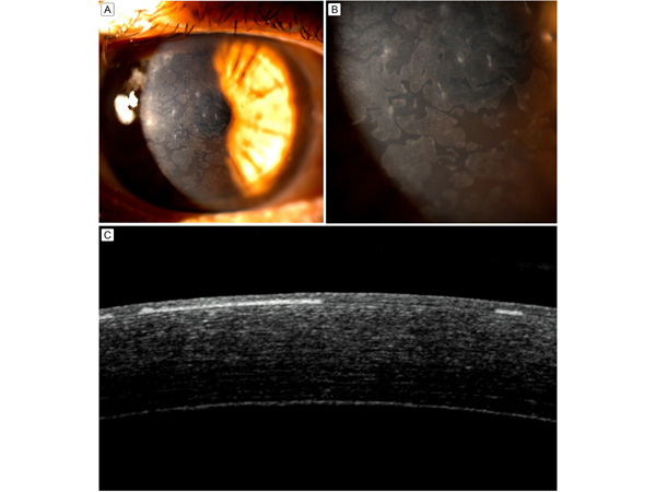
A 60-year-old woman presented at Sankara Eye Hospital, Guntur, Andhra Pradesh,India, with foreign body sensation and watering in her right eye. She had undergone cataract surgery 6 months earlier. Medical history included diabetes and hypertension, for which she was taking medications. On examination, best-corrected visual acuity was 6/24 in the right eye and 6/12 in the left eye. Slit-lamp examination showed multiple maplike subepithelial lesions with well demarcated margins spread diffusely on the cornea, with dispersed filaments (A, B). Her eye had a clear cornea, with senile cataract. Anterior segment optical coherence tomography (AS-OCT) showed well defined hyper-reflective lesions limited to the basement membrane (C). Dry eye evaluation was normal. A diagnosis of unilateral map-dot dystrophy was made based on clinical morphology and confirmed by AS-OCT.
Downloads
Article Details

This work is licensed under a Creative Commons Attribution-NonCommercial-NoDerivatives 4.0 International License.