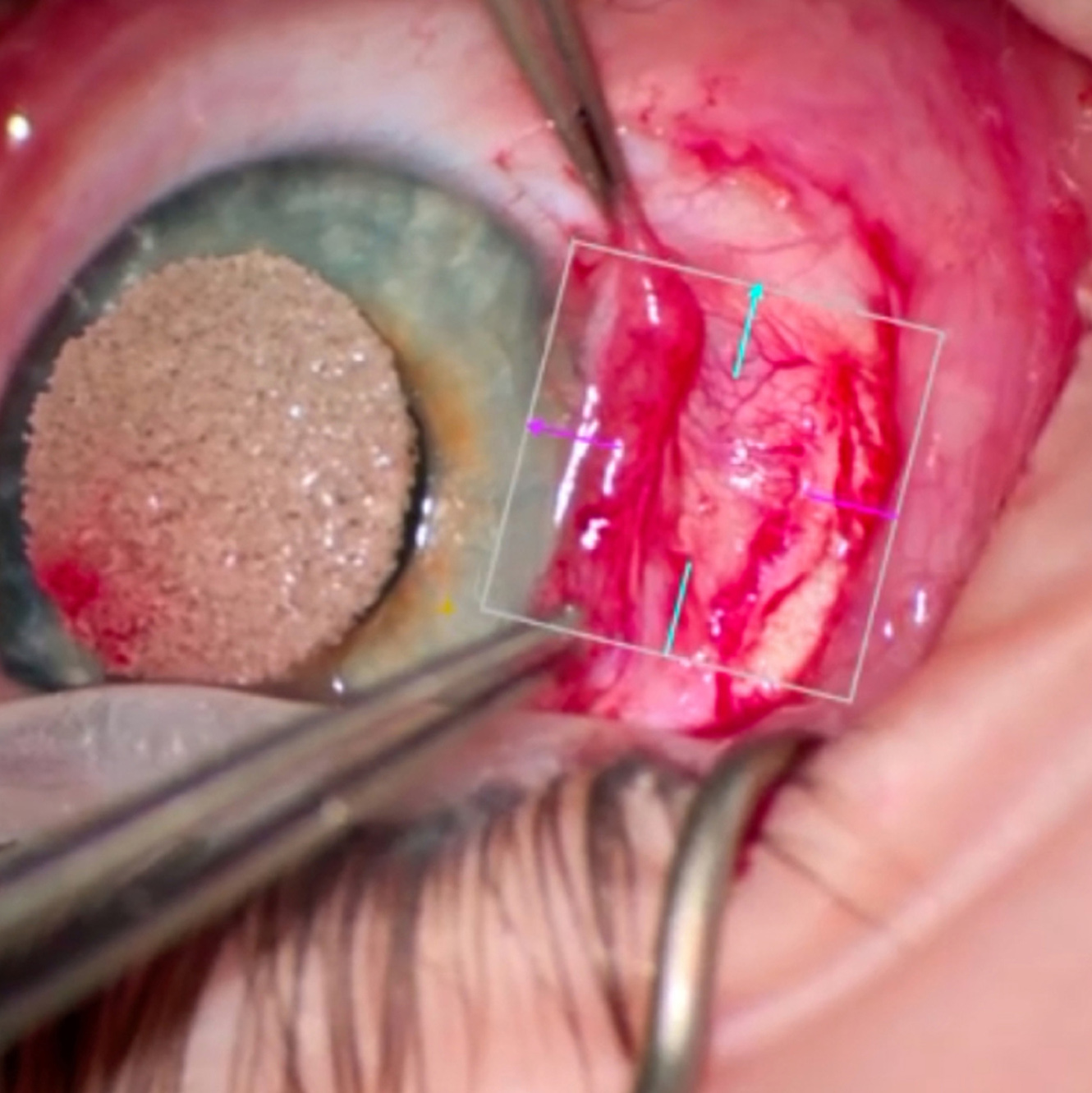
Excision of an intrascleral cyst guided by anterior segment optical coherence tomography
Main Article Content
Abstract
A 4-year-old girl presented with an enlarging, congenital, intrascleral cyst of the left eye. Intraoperative anterior segment optical coherence tomography was used to visualize and to assess the extent of the cyst, facilitating safe excision. The cyst was completely removed, and the defect was covered with an amniotic membrane graft, with a good outcome.
Downloads
Article Details

This work is licensed under a Creative Commons Attribution-NonCommercial-NoDerivatives 4.0 International License.
References
Akbaba M, Hacıyakupoğlu G, Uğuz A, Karslıoğlu S, Karcıoğlu Z. Congenital intrascleral cyst. Clin Ophthalmol 2011;5:583-5. DOI: https://doi.org/10.2147/OPTH.S19789
Mahmood MA, Awad A. Congenital sclerocorneal epithelial cyst. Am J Ophthalmol 1998;126:740-1. DOI: https://doi.org/10.1016/S0002-9394(98)00128-7
Liakos GM. Intracorneal and sclerocorneal cysts. Br J Ophthalmol 1978;62:155-8. DOI: https://doi.org/10.1136/bjo.62.3.155
Soni T, Das S. Natural course of congenital corneoscleral cyst: 10-year follow-up. Indian J Ophthalmol 2020;68:2217-8. DOI: https://doi.org/10.4103/ijo.IJO_206_20
Titiyal JS, Kaur M, Nair S, Sharma N. Intraoperative optical coherence tomography in anterior segment surgery. Surv Ophthalmol 2021;66:308-26. DOI: https://doi.org/10.1016/j.survophthal.2020.07.001


