Multiple subretinal fluid blebs after successful scleral buckling surgery
Article Sidebar
Main Article Content
Abstract

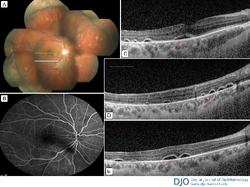
A 32-year-old man presented at Sankara Nethralaya, Kolkata, with metamorphopsia in his right eye, which began 3 months after he had undergone surgery for retinal detachment and 1 week before presenting at our clinic. Best-corrected visual acuity in his right eye was 20/30. Anterior segment examination was unremarkable. Color fundus photography showed that the retina was attached, though there was a buckle effect noted in the periphery (A). Spectral domain optical coherence tomography (SD-OCT) showed multiple subretinal fluid (SRF) blebs in the subfoveal region and the inferior part of the retina (C-E), with no leakage on fundus fluorescein angiography in the corresponding regions (B). (Green, white, and red lines indicate OCT scanning lines.) Multiple SRF blebs can be found after successful treatment of primary rhegmatogenous retinal detachment. They may occur in a previously detached retina during the postoperative period after successful surgery. SRF accumulation can develop several days to weeks after complete retinal reattachment as a consequence of scleral buckling, with or without cryotherapy. Various mechanisms have been described for SRF accumulation, including breakdown of the blood-retinal barrier as a result of surgery allowing an excessive quantity of protein to enter SRF and surgical trauma to the retinal pigment epithelium–Bruch membrane complex. These multiple SRF pockets are usually self-resolving. Fluorescein angiography helps in differentiating this condition from other causes of SRF, such as central serous chorioretinopathy or Vogt-Koyanagi-Harada disease, where leakage may be expected.
Downloads
Article Details

This work is licensed under a Creative Commons Attribution-NonCommercial-NoDerivatives 4.0 International License.