A 26-year-old man with a blind spot in his left eye
Article Sidebar
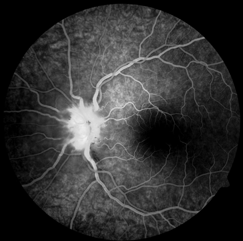
Main Article Content
Abstract
A 26-year-old white man presented with a 4-day history of a scotoma in the left eye only. He characterized the visual field defect as greenish and fixed in a superotemporal, paracentral location. The visual change was sudden in onset and was not associated with other ocular complaints or headache. Past ocular history was remarkable only for a diagnosis of herpes simplex keratitis in the involved eye. One episode had occurred 13 years before. It was treated with trifluridine and was not complicated by scarring or recurrence. Past medical history was unremarkable. Social history was negative for antecedent travel, illicit drugs, or trauma, but was positive for cat exposure. Review of systems was negative.
Downloads
Article Details

This work is licensed under a Creative Commons Attribution-NonCommercial-NoDerivatives 4.0 International License.




References
Davis JL, Madow B, Cornett J, et al. Scale for photographic grading of vitreous haze in uveitis. Am J Ophthalmol 2010;150:637-41.e1.
Cunningham ET Jr. What diseases should I consider in a patient with neuroretinitis? In Foster CS, Hinkle DM, Opremcak EM, eds. Curbside Consultation in Uveitis. Thorofare, NJ: Slack Book Incorporated; 2012:103-6.
Folk JC, Lobes LA. Presumed toxoplasmic papillitis. Ophthalmology 1984;91:64-7.
Gess A, Wender J, Jumper JM, Cunningham ET Jr. The red eye from Rio. EyeNet, May 2011:1-2.
Purvin V, Sundaram S, Kawasaki A. Neuroretinitis: review of the literature and new observations. J Neuroophthalmology, 2011;31:58-68.
Ray S, Gragoudas E. Neuroretinitis. Int Ophthalmol Clin 2001;41:83-102.
Holland GN. Reconsidering the pathogenesis of ocular toxoplasmosis. Am J Ophthalmol 1999;128:502-5.
Roberts F, McLeod R. Pathogenesis of toxoplasmic retinochoroiditis. Parasitol Today 1999;15:51-7.
Smith JR, Cunningham ET Jr. Atypical presentations of ocular toxoplasmosis. Curr Opin Ophthalmol 2002;13:387-92.
Eckert GU, Melamed J, Menegaz B. Optic nerve changes in ocular toxoplasmosis. Eye 2007;21:746-51.
Bonfioli AA, Orefice F. Toxoplasmosis. Semin Ophthalmol 2005;20:129-41.
Waragai M, Takaya Y, Hayashi M. [A case of toxoplasmic chorioretinitis and meningoencephalitis in an immunocompetent adult]. Rinsho Shinkeigaku 1995;35:559-62.
Papadia, M., R. Aldigeri, and C.P. Herbort, The role of serology in active ocular toxoplasmosis. International Ophthalmology 2011;31:461-5.
Rothova A. Ocular manifestations of toxoplasmosis. Curr Opin Ophthalmol 2003;14:384-8.
Commodaro AG, Belfort RN, Rizzo LV, et al. Ocular toxoplasmosis—an update and review of the literature. Oswaldo CruzMem Inst 2009;104:345-50.