Post-traumatic phacotopic glaucoma
Article Sidebar
Main Article Content
Abstract
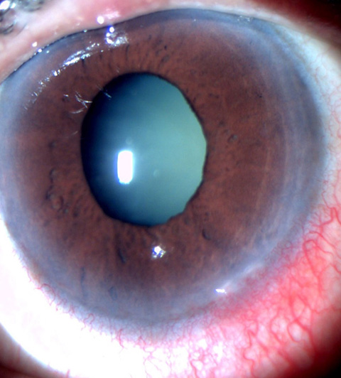
A 62-year-old man presented at Guru Nanak Eye Centre, New Delhi, with unilateral painless diminution of vision in his left eye following accidental blunt trauma by elbow. On examination, visual acuity in the eye was 6/60 (logMAR 1.00), with brisk pupillary reflex. Anterior segment evaluation showed a clear cornea with shallow anterior chamber (Van Herick grade1) and angle closure in all quadrants on gonioscopy. Dilated evaluation revealed 6 clock hours of anterior and temporally subluxated cataractous lens, from 6 to 12 o’clock, with loss of zonules in the subluxated area. There was no gross phacodonesis, no inflammatory reaction nor vitreous in anterior chamber, and intraocular pressure was 26 mm Hg in that eye. Posterior segment findings were unremarkable, with a healthy neuroretinal rim. The right eye had no significant abnormality. Anterior segment optical coherence tomography showed an anteriorly subluxated cataractous lens with peripheral iridocorneal contact. Gonioscopy showed closed inferior, superior, temporal, and nasal angles. A diagnosis of phacotopic glaucoma was made and a prophylactic peripheral iridotomy with frequency doubled Nd:YAG laser was performed to prevent pupillary block. The patient was scheduled for intracapsular cataract extraction with scleral-fixated intraocular lens under the cover of ocular hypotensive medications. Following surgery, the patient had an acceptable visual outcome of 6/12 (logMAR 0.30), with normal intraocular pressures at the end of 6 months’ follow-up.
Downloads
Article Details

This work is licensed under a Creative Commons Attribution-NonCommercial-NoDerivatives 4.0 International License.