Volcanic foveal herniation
Article Sidebar
Main Article Content
Abstract
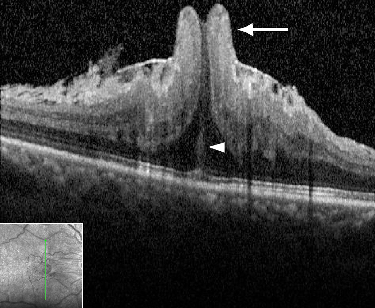
A 61-year-old man presented with progressive blurry vision and metamorphopsia of the right eye. Visual acuity was 20/250. Examination revealed an epiretinal membrane (ERM) with a concentric ring of foveal tissue prolapse. Optical coherence tomography (OCT) showed the fovea protruding through and above the ERM, demonstrating the severe foveal herniation (arrow, A). Foveal herniation has been recently described.1–3 The pathogenesis of foveal herniation is still unknown, but likely, at least in part, a result of centripetal ERM contraction and the characteristically thin pre-foveolar internal limiting membrane (ILM).4–6 In this particular case of severe volcano-like herniation, there may be secondary stretching of presumed Müller cell processes also (arrowhead, A). The patient underwent successful vitrectomy with ERM and ILM peeling, resulting in restoration of the foveal anatomy and improvement in vision to 20/40 (B).
Downloads
Article Details

This work is licensed under a Creative Commons Attribution-NonCommercial-NoDerivatives 4.0 International License.