Macular schisis associated with optic disc coloboma
Article Sidebar
Main Article Content
Abstract
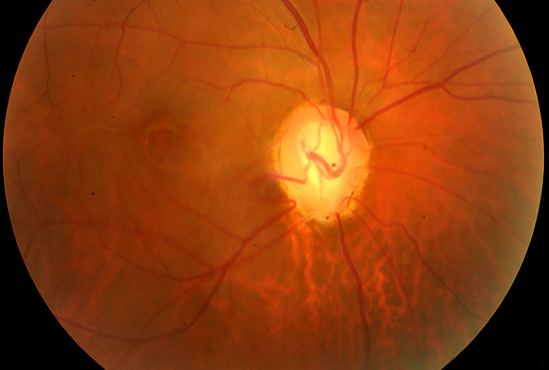
A 44-year-old man presented at the Aravind Eye Hospital with decreased vision and metamorphopsia in his right eye of 5 months’ duration. On examination, best-corrected visual acuity was 20/40. Fundus examination of the right eye revealed that the optic disc had a bowl-shaped excavation inferonasally, suggestive of a coloboma (A). The macula had a schitic, cystoid appearance. Enhanced-depth optical coherence tomography (OCT) of the right eye through the optic disc and macula shows schisis with neurosensory detachment (B). This schisis is present in all layers but predominantly involves the outer plexiform layer. The communication between the perineural space and subretinal space is seen (black arrow). Enhanced-depth OCT through the optic disc shows a discontinuity suggestive of the coloboma (C). The scleral defect in the optic disc due to the coloboma creates an anatomical connection between the intra- and extraocular spaces. Fluctuations of intraocular and intracranial pressure directs fluid through the defect and into the retina, resulting in schisis. Because visual acuity was good, observation was advised, with regular review every 3 months. The patient was lost to follow-up.
Downloads
Article Details

This work is licensed under a Creative Commons Attribution-NonCommercial-NoDerivatives 4.0 International License.