Granulomatosis with polyangiitis presenting as central retinal artery occlusion
Article Sidebar
Main Article Content
Abstract
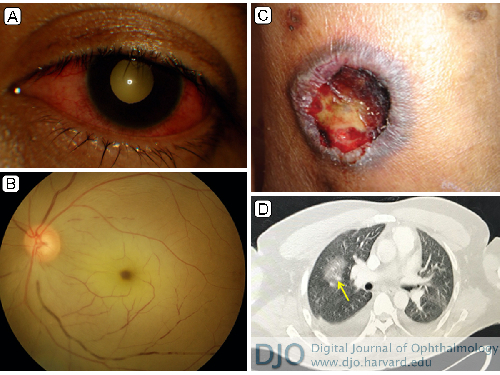
A 42-year-old man presented at the eye clinic of Penang Hospital, Malaysia, with bilateral eye redness and discomfort of 2 weeks’ duration. Two days prior to presenting, he experienced a sudden deterioration of vision in his left eye. On examination, visual acuity was 6/9 in the right eye and light perception in the left eye, with a left relative afferent pupillary defect. There was bilateral, nongranulomatous anterior uveitis (A, top [left eye]), with central retinal artery occlusion of the left eye (A, bottom); right eye fundus examination was unremarkable. Blood investigations showed markedly elevated C-reactive protein and the presence of cytoplasmic antineutrophil cytoplasmic antibodies (c-ANCA). Systemic examination revealed bilateral lower limb cutaneous ulcerations (B) consistent with pyoderma gangrenosum. Contrast-enhanced computed tomography showed pansinusitis, with multiple lung nodules and consolidation (C, arrow). The patient achieved remission after being treated with systemic steroids and 6 cycles of cyclophosphamide. Although uncommon, ocular signs may be the initial presenting signs of a multisystem vasculitis, such as polyangiitis with granulomatosis, as is the diagnosis in this case.
Downloads
Article Details

This work is licensed under a Creative Commons Attribution-NonCommercial-NoDerivatives 4.0 International License.