Acute posterior multifocal placoid pigment epitheliopathy
Article Sidebar
Main Article Content
Abstract
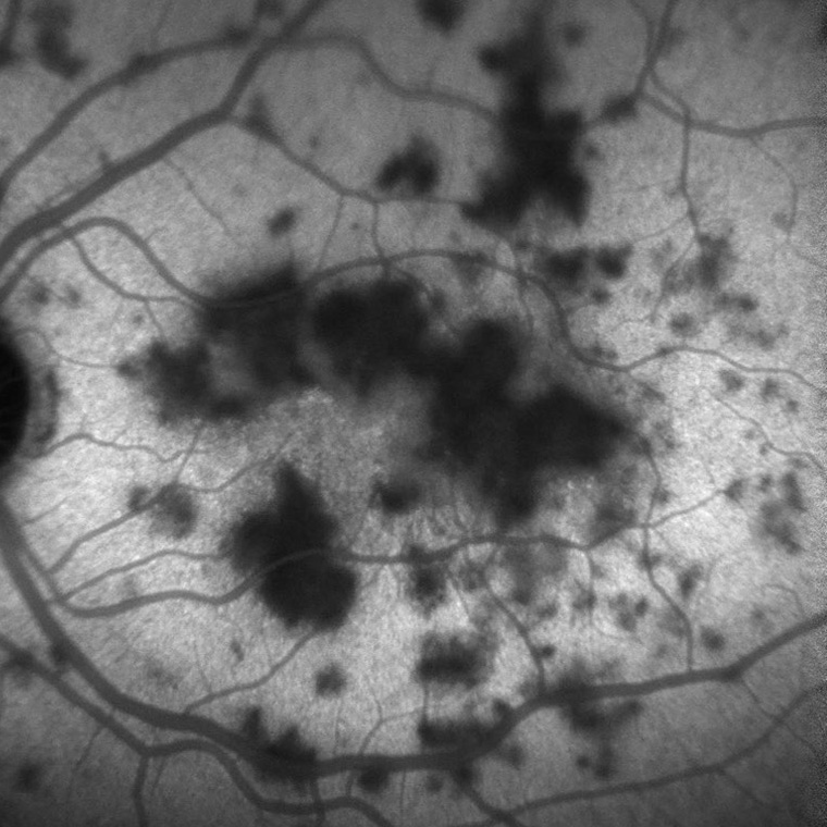
Acute posterior multifocal placoid pigment epitheliopathy (APMPPE) is a rare but important uveitic entity that can be associated with neurologic complications, including cerebral vasculitis, headache, and stroke. A 31-year-old man presented at Greenlane Clinical Centre, Auckland, New Zealand, with blurred vision. On examination, visual acuity was 20/40 in the right eye and 20/32 in the left eye. Both eyes had similar findings. Fundus photography of the left eye demonstrated multiple creamy placoid lesions at the posterior pole (A). On indocyanine green angiography (B), these placoid lesions were hypofluorescent and more numerous than on clinical examination. On fundus fluorescein angiography, the placoid lesions were hypofluorescent in the early phase (C) and became hyperfluorescent in the later phases (D). Optical coherence tomography revealed areas of hyper-reflectivity in the lesions at the level of the outer retina, involving all layers from the outer plexiform layer to the retinal pigment epithelium (E). These findings are consistent with APMPPE.
Downloads
Article Details

This work is licensed under a Creative Commons Attribution-NonCommercial-NoDerivatives 4.0 International License.
References
not required for images