Progressive enlargement of diffuse iris melanoma
Article Sidebar
Main Article Content
Abstract
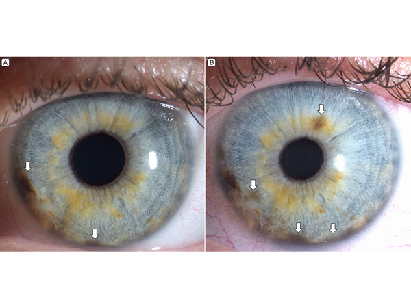
A 36-year-old white woman presented at the Ocular Oncology Service, Wills Eye Hospital, with an iris freckle in the left eye since childhood showing possible recent enlargement. On slit-lamp examination of the left eye (A), the flat, pigmented iris lesion appeared discohesive and extended from the 6:00 to 9:30 meridians in the iris root and into the angle (arrows). By ultrasound biomicroscopy, the lesion was 1.2 mm in thickness and without ciliary body involvement. Observation was advised. At 4 months’ follow-up (B), a new focal area of pigmentation along the 1:00 meridian and increased pigmentation inferonasally were noted (arrows). Gonioscopy showed heavy pigmentation in the angle inferonasally; ultrasound biomicroscopy was stable. Fine-needle aspiration biopsy verified the diagnosis of diffuse iris melanoma. Treatment with iodine-125 plaque radiotherapy was provided. This patient demonstrated 4 of 6 risk factors for nevus growth into melanoma, including young age, inferior clock hour, diffuse configuration, and feathery margins. Support provided in part by the Eye Tumor Research Foundation, Philadelphia, PA (CLS).
Downloads
Article Details

This work is licensed under a Creative Commons Attribution-NonCommercial-NoDerivatives 4.0 International License.