Macular oxalosis associated with choroidal neovascularization in childhood
Article Sidebar
Main Article Content
Abstract
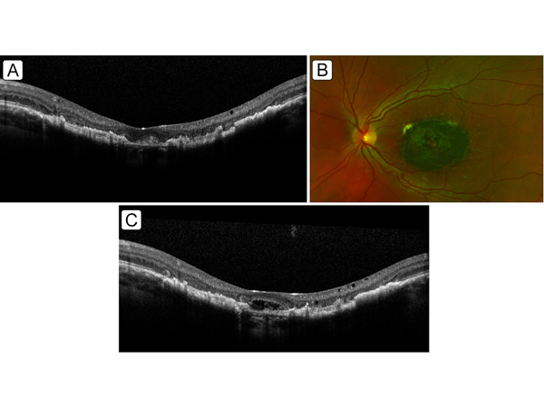
A 13-year-old boy with a diagnosis of infantile hyperoxaluria type 1 (PH1) and a history of hepatorenal transplant presented at the Casey Eye Institute, Oregon Health & Science University, with recent-onset of metamorphopsia in his left eye. Before anti-VEGF therapy, cross-sectional optical coherence tomography (OCT) demonstrated crystal deposits, appearing as subretinal hyperreflective lesions in both eyes, as well as focal subretinal fluid and intraretinal cystoid spaces in the left eye, suggestive of a choroidal neovascular (CNV) membrane (A). Fundus photography showed a dense annular area of macular pigmentation sparing the fovea in both eyes (B). He was treated with three monthly bevacizumab injections in the left eye, with subsequent resolution of the fluid (C) and improvement in visual acuity from 20/50 to 20/25. Oxalosis typically demonstrates metaplasia of the retinal pigment epithelium (RPE), which may be an inflammatory response to deposition of crystalline oxalates below the RPE. This case highlights the possibility of CNV as a treatable complication of this ultrarare disease. The authors have been funded in part by the Coordenação de Aperfeiçoamento de Pessoa de Nível Superior – Brasil (CAPES) finance code 001 (MMP) and by grants R01EY19474, EY031331, and P30EY10572 from the National Institutes of Health, Bethesda, MD, and by unrestricted departmental funding and a Career Development Award (JPC) from Research to Prevent Blindness, New York, NY (JPC).
Downloads
Article Details

This work is licensed under a Creative Commons Attribution-NonCommercial-NoDerivatives 4.0 International License.