Serous choroidal effusion with bridging vortex vessel vein
Article Sidebar
Main Article Content
Abstract
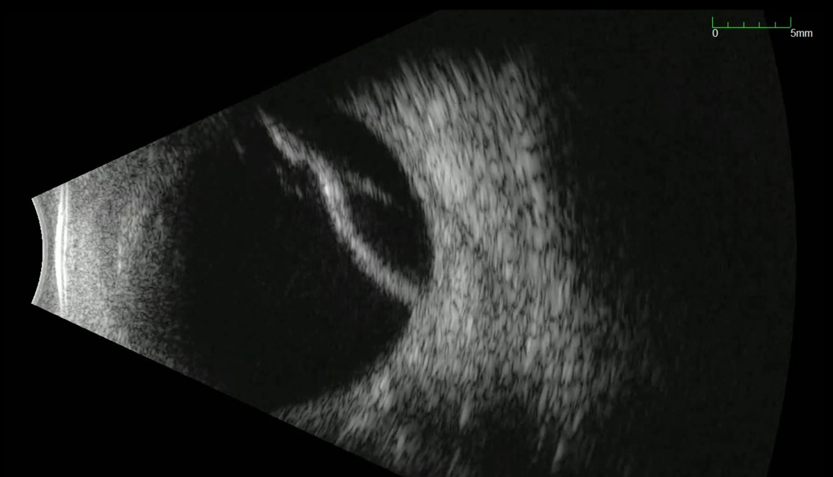
A 78-year-old male presented with sudden decrease in vision. He had a history of recent right eye spontaneously dislocated IOL that was treated with pars plana vitrectomy, IOL explanation and ACIOL placement. On post-operative day 10, he had sudden vision loss and was referred to our institution for repair of a presumed retinal detachment diagnosed by an outside ultrasound. His visual acuity was hand-motions, and the intraocular pressure was 4mmHg. The ACIOL was in good position and vitreous hemorrhage (VH) obscured the posterior pole. Dynamic B-scan ultrasonography revealed a mobile, irregular, hyperechoic membrane with an underlying bridging structure indicative of a serous choroidal effusion (SCE). Though normally SCE appear as smooth and immobile dome-shaped structures on ultrasound, the bridging vessel in this case changed the appearance and behavior of the SCE with eye movements. Recognition of the bridging vessel, in addition to the thickness of the echogenic structure, aided in the conclusion of SCE and not retinal detachment. The mainstay of treatment for hypotony-induced SCE is restoration of normal intraocular pressure. In this case, the ACIOL scleral wound likely leaked and resulted in acute hypotony, VH and SCE. On exam, the wound was seidel negative, and therefore observation was indicated. Three days after presentation, the IOP increased to 10, and the serous choroidal detachment receded. The final visual acuity at month 1 was 20/40 and the retina remained attached. This story showcases a unique ultrasonographic view that helps distinguish a serous choroidal effusion from a retinal detachment on ultrasound.
Downloads
Article Details

This work is licensed under a Creative Commons Attribution-NonCommercial-NoDerivatives 4.0 International License.