Multimodal imaging in a case of congenital intertwined retinal vessels
Article Sidebar
Published:
Jul 27, 2023
Keywords:
retina, Coats disease
Main Article Content
Abstract
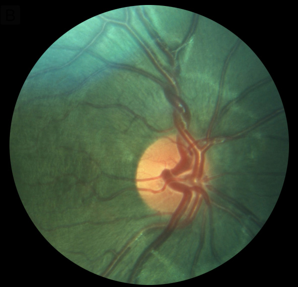
A 7-year-old boy presented at Guru Nanak Eye Centre, New Delhi, with ringlike exudation posteriorly and superonasally, superotemporally, and inferonasally in his right eye (A); his left eye was normal. He was diagnosed with Coats disease. On examination, he was found to have intertwining of the central retinal artery and vein in the right eye, close to the disc (B), which could be better appreciated on fluorescein angiography (C). The child underwent laser barrage for Coats disease and has been followed closely.
Downloads
Download data is not yet available.
Article Details
How to Cite
1.
Bhattacharya S, Bansal P, Thakar M. Multimodal imaging in a case of congenital intertwined retinal vessels. Digit J Ophthalmol. 2023;29(3). Accessed July 28, 2026. https://djo.harvard.edu/index.php/djo/article/view/440
Issue
Section
Images & Videos

This work is licensed under a Creative Commons Attribution-NonCommercial-NoDerivatives 4.0 International License.