Persistence of rifabutin-related peripheral circumferential corneal endothelial deposits
Article Sidebar
Main Article Content
Abstract
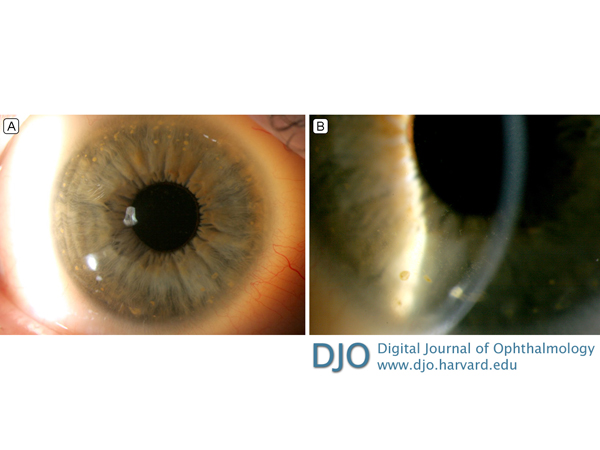
A 38-year-old man was referred to the cornea clinic at Cambridge University Hospitals for an incidental finding of bilateral corneal endothelial deposits. Past medical history included tuberculous cervical lymphadenitis and Crohn’s disease (CD) with terminal ileitis at 12 years of age, right hemicolectomy, and enteropathic arthritis. The patient was asymptomatic, with 20/20 unaided visual acuity bilaterally and normal intraocular pressures. Anterior segment examination revealed multiple, bilateral, peripheral, large, yellow corneal endothelial deposits, circumferentially distributed, sparing the central cornea. Additionally, there were fine, stellate, yellow deposits, which also spared the central cornea but extended further toward the mid-cornea. The distribution of larger deposits can be seen in the scleral scatter image (A); fine details of both the larger and the stellate deposits are visible in the slit-lamp image (B). Occasional anterior chamber inflammatory cells were observed bilaterally. Funduscopy was unremarkable. The patient reported a concurrent CD flare-up. A diagnosis of anterior uveitis with keratic precipitates (KPs) associated with CD relapse was made initially. However, the atypical location and appearance of the deposits prompted further questioning. It transpired that he had received rifabutin-clarithromycin treatment for CD 24 years previously. Rifabutin-related corneal endothelial deposits are usually asymptomatic and can be overlooked or misinterpreted as KPs, particularly in the context of systemic autoimmune disease. They have been reported to persist for up to 10 years after cessation of treatment. Our case demonstrates that they can persist for at least 22 years. The characteristic distribution and features can be considered pathognomonic. Rifabutin use can also be associated with lenticular opacities and retinal dysfunction. We do not know in this case whether the mild anterior uveitis was related to rifabutin therapy or CD.
Downloads
Article Details

This work is licensed under a Creative Commons Attribution-NonCommercial-NoDerivatives 4.0 International License.