Proboscis lateralis in a newborn
Article Sidebar
Main Article Content
Abstract
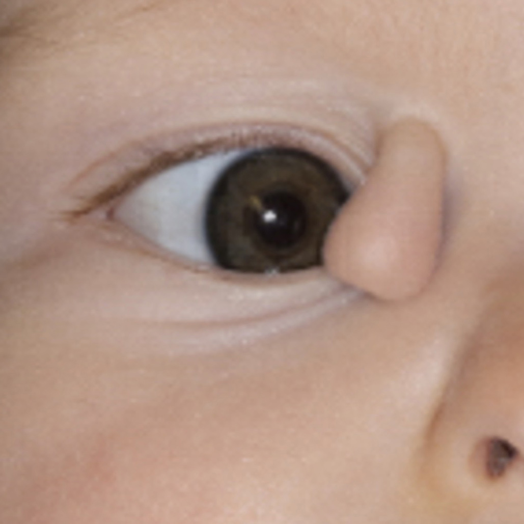
A 2-week-old girl, born at full-term following an unremarkable pregnancy, presented at the Oculoplastics Clinic, Byers Eye Institute, for evaluation of a soft, tubelike appendage extending from the region above the right medial canthus accompanied by ipsilateral hypoplastic naris (A). The tubular appendage, measuring 2.75 cm long and 1 cm wide, was present at birth. Magnetic resonance imaging revealed a soft-tissue mass with a bony fragment at its base directly anterior to the trochlea along with subcutaneous extension (B, yellow asterisk) but no communication with other orbital, nasal, oral, or intracranial structures. A diagnosis of proboscis lateralis was made. Surgical treatment of proboscis laterals is individualized to the type of presentation and imaging findings. Surgical excision of the tubular medial canthal lesion, which can subsequently be used for reconstruction of significant nasal defects, is typically recommended, and management may benefit from a multidisciplinary approach involving ophthalmology, otolaryngology, and craniofacial plastic surgery. Operative repair for this patient, planned for 11 months of age, involved complete excision of the proboscis lateralis and layered closure. There were no intra- or postoperative complications, and the patient’s postoperative course has been unremarkable, with satisfactory cosmesis at 1 week and 2 months’ follow-up.
Downloads
Article Details

This work is licensed under a Creative Commons Attribution-NonCommercial-NoDerivatives 4.0 International License.