Bilateral progressive choroidal osteomas causing vision impairment
Article Sidebar
Main Article Content
Abstract
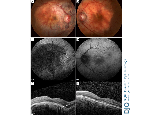
A 27-year-old healthy woman presented at the Royal Victorian Eye and Ear Hospital with bilateral gradual reduction in vision over several months. On examination, best-corrected visual acuity was 6/15 in the right eye and 6/12 in the left eye. Fundus examination revealed elevated, subretinal, pale-orange lesions with scalloped margins encompassing the posterior poles, more extensive in right eye (A, B). B-scan ultrasonography revealed bilateral, focal, hyperechoic choroidal signals with posterior shadowing. Fundus autofluorescence of the right eye demonstrated large areas of hypoautofluorescence indicating decalcification and retinal pigment epithelium (RPE) atrophy, with surrounding hyperautofluorescence suggesting RPE disturbance (C, D). Optical coherence tomography (OCT) revealed elevated choroidal lesions (more prominent in right eye), with a lamellar and spongy appearance (E, F). Nasal to the right fovea was an area (E, arrow) of subtle intralesional hyper-reflectivity (suggestive of early decalcification) with overlying disruption of the RPE, ellipsoid zone, and external limiting membrane. The patient was diagnosed with bilateral progressive choroidal osteomas, with vision loss due to outer retinal disruption. Although benign, these rare ossifying tumors may result in vision loss through choroidal neovascularization, subretinal fluid, or atrophy of the RPE or outer retina. In contrast to calcified osteomas that appear to maintain intact photoreceptor and retinal anatomy, tumor decalcification (seen as hyper-reflective regions on OCT) is associated with atrophy of the RPE or outer retina and photoreceptor retraction corresponding with gradual vision impairment.
Downloads
Article Details

This work is licensed under a Creative Commons Attribution-NonCommercial-NoDerivatives 4.0 International License.