Bluish discoloration in the eye—nevus of Ota
Article Sidebar
Main Article Content
Abstract
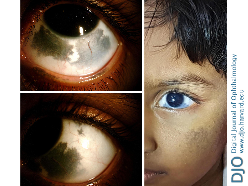
A 1-year-old child presented at the Rajan Eye Care Hospital for assessment of dark blue spots in the white part of the left eye since birth. The child was able to fix and follow. On examination, the left eye showed grayish-blue hyperpigmentation of the sclera, most prominent inferiorly. There was also bluish discoloration of the ipsilateral facial skin over the cheek. On examination, the anterior segment of the right eye and posterior segments of both eyes were normal. The child has been under regular follow-up for 4 years, and visual acuity, intraocular pressure, and fundus findings have remained stable during this time. Slit-lamp images of the left eye and a clinical photograph of the face at the most recent follow-up are shown. Nevus of Ota, or oculomucodermal melanocytosis, is a benign condition that causes ipsilateral hyperpigmentation that may involve the eyelids, conjunctiva, sclera, uvea, orbit, and the face in the distribution of the ophthalmic and maxillary divisions of the trigeminal nerve. Less common sites include the meninges, palate, and tympanic membrane. It usually presents at birth but can present later and usually remains unchanged throughout life. Regular follow-up is required, because there is a risk of glaucoma and malignant ocular or cutaneous melanoma.
Downloads
Article Details

This work is licensed under a Creative Commons Attribution-NonCommercial-NoDerivatives 4.0 International License.