Fluorescein angiography of a retinal venous malformation
Article Sidebar
Main Article Content
Abstract
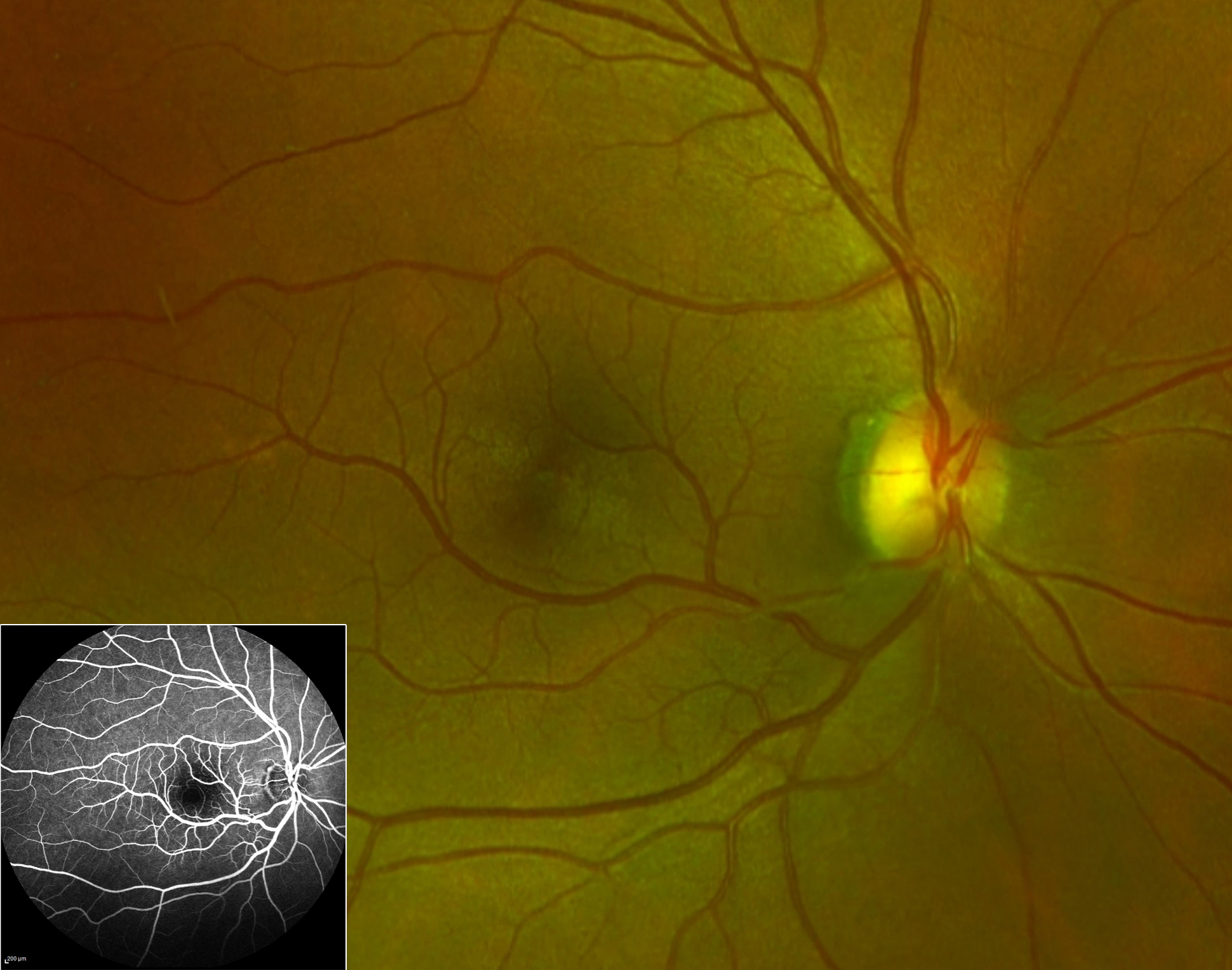
A 65-year-old woman who presented to Ben Taub General Hospital for routine evaluation was found on examination to have a retinal venous malformation (RVM), also referred to as congenital retinal macrovessel, in her right eye. The left eye was normal. Fundus photography of the right eye (A) revealed a dilated anomalous retinal blood vessel traversing the horizontal raphe across the central macula. Fluorescein angiography (B) highlighted the inferotemporal retinal venous malformation (arrows) and its crossing of the superotemporal vasculature. RVMs are aberrant retinal vessels (typically veins), with a vascular distribution that crosses the horizontal raphe. Often unilateral and asymptomatic, RVMs have been associated with retinal macroaneurysm, branch retinal artery occlusion, retinal telangiectasia, and even brain venous malformation. While they are considered arteriovenous malformations by some, the site of arteriovenous communication is virtually impossible to localize on fluorescein angiography.
Downloads
Article Details

This work is licensed under a Creative Commons Attribution-NonCommercial-NoDerivatives 4.0 International License.