Conjunctival choristoma
Article Sidebar
Main Article Content
Abstract
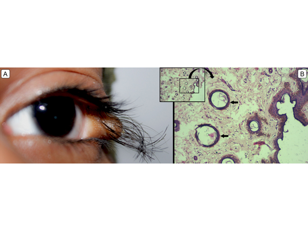
A 10-year-old girl presented at Sankara Nethralaya, Chennai, India, for evaluation of a painless, progressive mass in her left eye since birth. Her birth history and family history were unremarkable. Uncorrected visual acuity in each eye was 20/20. Examination revealed a pinkish-brown elevated mass with hairs at the lateral canthus of the left eye (A). She was orthophoric, with unrestricted ocular movements. The rest of the ocular and physical examination revealed no other abnormalities. Choristomas are congenital malformations consisting of normal elements in an abnormal location. Conjunctival dermoids are simple choristomas, because they contain one germ cell layer lined by a conjunctival epithelium with deeper dermal elements, including hair follicles (arrow) and sebaceous glands involving the bulbar or limbal conjunctiva (B). The surface can be keratinized and the dermoid usually occurs in the inferotemporal quadrant. A complex choristoma contains tissues derived from two germ layers, such as lacrimal tissue and cartilage. Conjunctival dermoids can be sporadic, or they can be associated with Goldenhar syndrome. Cosmesis and astigmatism are common complaints that warrant treatment. The management of a conjunctival choristoma includes observation, if the lesion is cosmetically acceptable, and visually asymptomatic. Anterior segment optical coherence tomography can assist in judging the depth of involvement. It can be managed by careful excision or debulking of the anterior portion of the lesion through a conjunctival forniceal approach, with mucous membrane or autologous conjunctival graft reconstruction and lateral canthal repair. The possibility of incomplete removal, diplopia, ptosis, and resultant scarring should be discussed with the patient beforehand.
Downloads
Article Details

This work is licensed under a Creative Commons Attribution-NonCommercial-NoDerivatives 4.0 International License.