Whiplash-like movement of persistent fetal vasculature
Article Sidebar
Main Article Content
Abstract
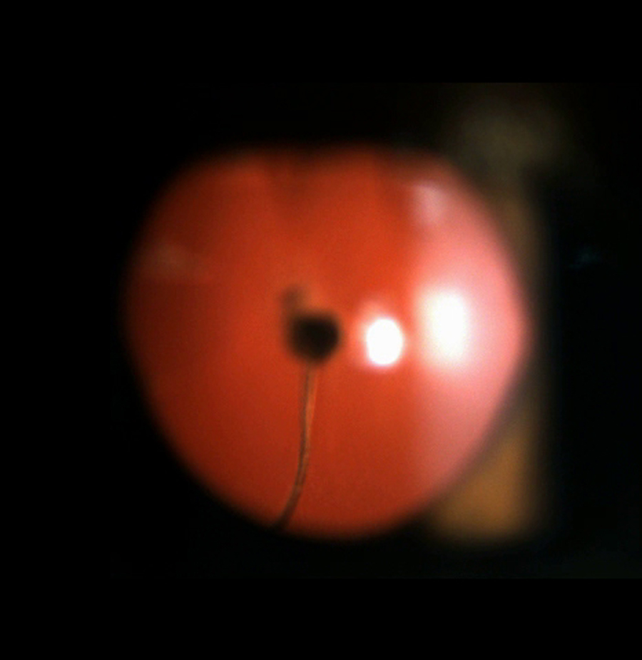
A 16-year-old young man presented at the Dr. Rajendra Prasad Centre for Ophthalmic Sciences, New Delhi, with diminished vision in his left eye of 4 years' duration. On examination, best-corrected visual acuity was 6/6 in the right eye and 3/60 (−1.00 −0.50 ×100) in the left eye. Corneal diameters were normal in both eyes. In the left eye there were several wedge-shaped cortical lenticular opacities and a posterior polar cataract. A stalk connecting the cataract with the nasal side of the disc moved with a "whiplash" motion (Video). There was focal chorioretinal degeneration surrounding the optic disc and a posterior staphyloma. The right axial length of was 24.98 mm and the axial length of the left eye was 23.81 mm. After lens aspiration, posterior capsulorhexis, endocautery of the persistent fetal vascular stalk and anterior vitrectomy and placement of a multipiece intraocular lens in the capsular bag, the patient's visual acuity improved to 6/36.
Downloads
Article Details

This work is licensed under a Creative Commons Attribution-NonCommercial-NoDerivatives 4.0 International License.