Extraocular muscle belly enlargement with myotendinous sparing in thyroid-associated orbitopathy
Article Sidebar
Main Article Content
Abstract
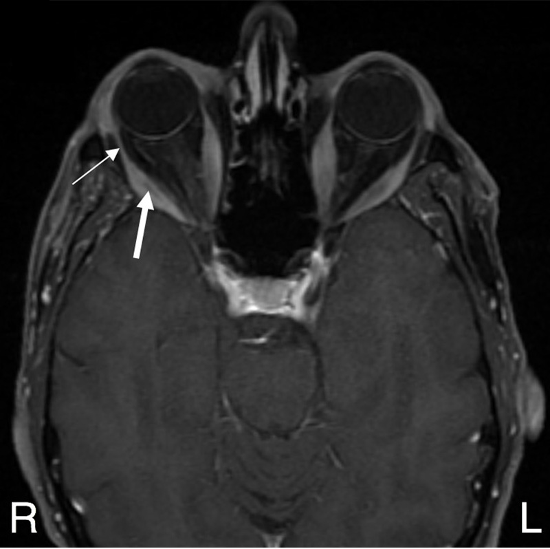
A 66-year-old man with Graves disease presented at the Yale New Haven Hospital for evaluation of worsening binocular diplopia. TSH levels were low (<0.005 μIU/mL), and thyroid peroxidase antibodies were elevated (555 IU/mL [normal, <35 IU/mL]). Coronal (A) and axial (B) T1-weighted magnetic resonance imaging with contrast showed bilateral enlargement of inferior, medial, superior, and lateral rectus muscle bellies (B, thick arrow), with sparing of the myotendinous junctions (B, thin arrow), consistent with thyroid-associated orbitopathy. Axial T2-weighted imaging (C) showed right globe anterior positioning measured from the interzygomatic line (normal, below 21 mm). Thyroid-associated orbitopathy affects 20%-50% of patients with Graves disease. It may be caused by autoantibody activation of orbital fibroblasts, causing inflammation and glycosaminoglycan deposition. The condition affects bilateral extraocular muscles 90% of the time; the inferior rectus muscle is most likely to be involved, followed by the medial rectus, superior rectus, lateral rectus, and oblique muscles.
Downloads
Article Details

This work is licensed under a Creative Commons Attribution-NonCommercial-NoDerivatives 4.0 International License.