Spontaneous hemorrhage from an iris vascular tuft
Article Sidebar
Main Article Content
Abstract
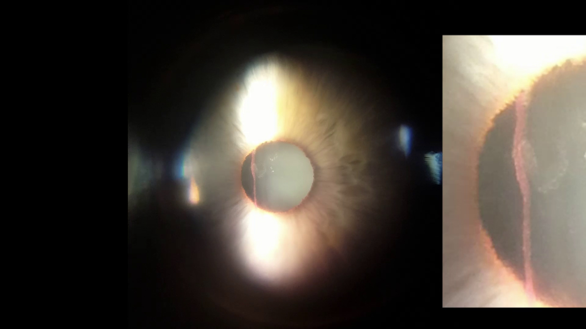
A 55-year-old woman presented at Royal Gwent Hospital, Newport, with blurry vision in her right eye on waking that morning. Later that day, she practiced Pilates, after which her vision drastically worsened. She denied any past medical history, recent trauma, or similar episodes previously. On examination that afternoon, her visual acuity had already improved subjectively and measured 6/4 in the right eye. Iris vascular tufts, or Cobb’s tufts, were noted bilaterally, from which there was an active hemorrhage from the right pupillary margin at the 11 o’clock position (video). A small amount of hyphema was seen, and the intraocular pressure (IOP) was 28 mm Hg in the right eye and 16 mm Hg in the left eye. A gonioscope was applied onto the right eye gently for 2 minutes, which halted the bleeding immediately and after removal of the gonioscope. Her blood pressure and blood glucose level were found to be within normal limits (126/80 mm Hg and 5.0 mmol/L, resp.). She was prescribed steroid and ocular antihypertensive eyedrops. On review 3 days later, her IOP was 16 mm Hg in the right eye. Cup-disc ratios were noted to be 0.2 bilaterally. Fundi appeared normal in both eyes. Topical antiglaucoma medication was discontinued then, and steroid eyedrops were tapered gradually and stopped after a week. She reported no further episodes over 2 months’ follow-up.
Downloads
Article Details

This work is licensed under a Creative Commons Attribution-NonCommercial-NoDerivatives 4.0 International License.