Corneal keloid
Article Sidebar
Main Article Content
Abstract
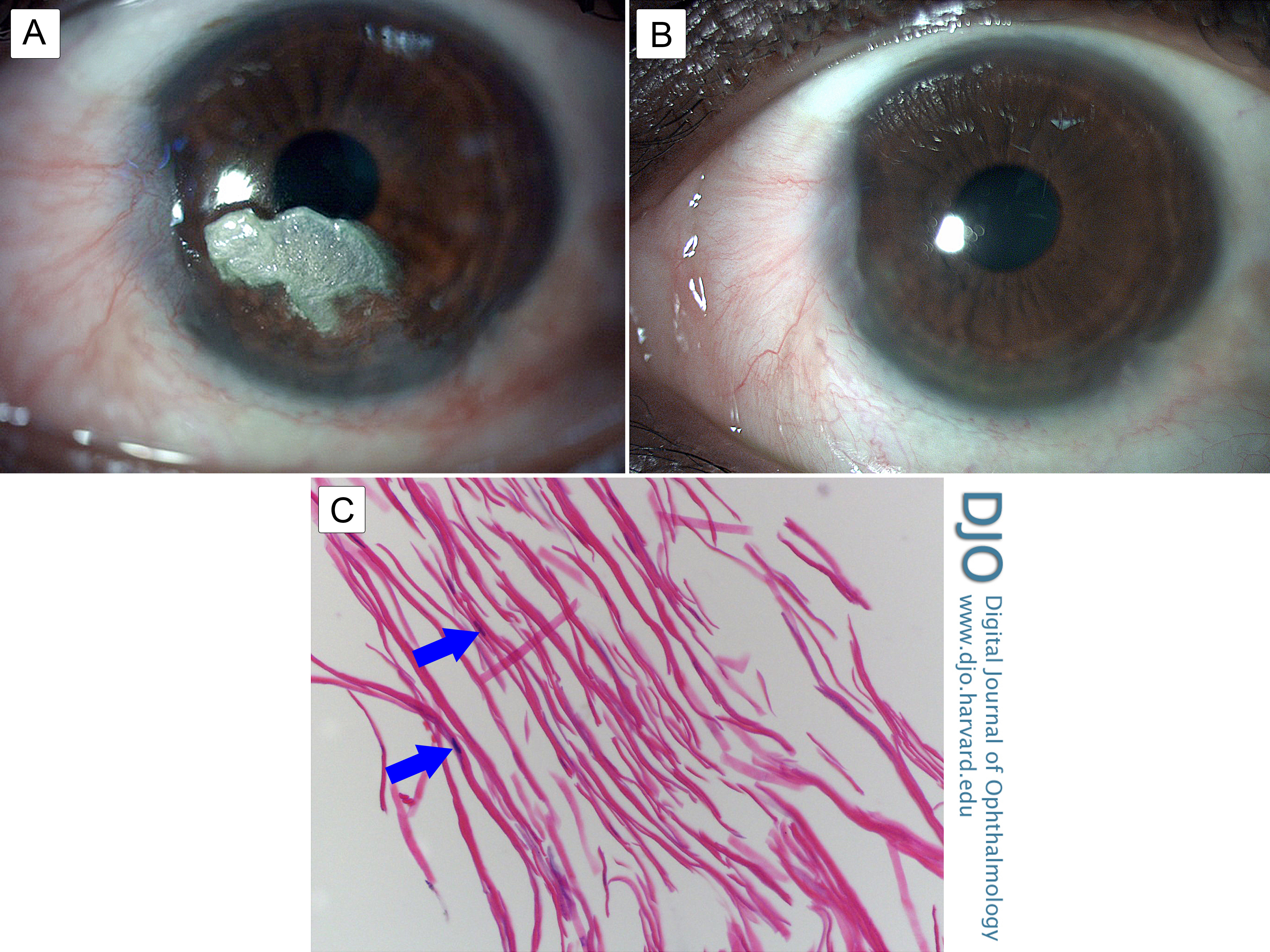
A 37-year-old woman presented at Kings County Hospital Center with a 6-month history of a slow-growing “white spot” in her left eye. She had undergone pterygium excision in the same eye 6 years before, with a small recurrence noted a year after excision. She also had a remote history of keloid formation on both earlobes after piercing. On examination, visual acuity was 20/30 in the right eye and 20/40 in the left eye. Slit-lamp biomicroscopy revealed a pearly, white lesion on the corneal epithelium with distinct margins and a smooth surface (A). The lesion was removed by superficial keratectomy, and a wet amniotic membrane graft was placed on the residual corneal stromal bed (B; postoperative week 3). Histopathology revealed a haphazard arrangement of fibroblasts (arrows) between collagen bundles, confirming the diagnosis of corneal keloid (C; hematoxylin-eosin, original magnification ×200). Four months after surgery, the patient’s visual acuity improved to 20/20, and there was no recurrence with 15 months’ follow-up.
Downloads
Article Details

This work is licensed under a Creative Commons Attribution-NonCommercial-NoDerivatives 4.0 International License.