Lobulated uveal melanoma with both pigmented and nonpigmented tumor
Article Sidebar
Main Article Content
Abstract
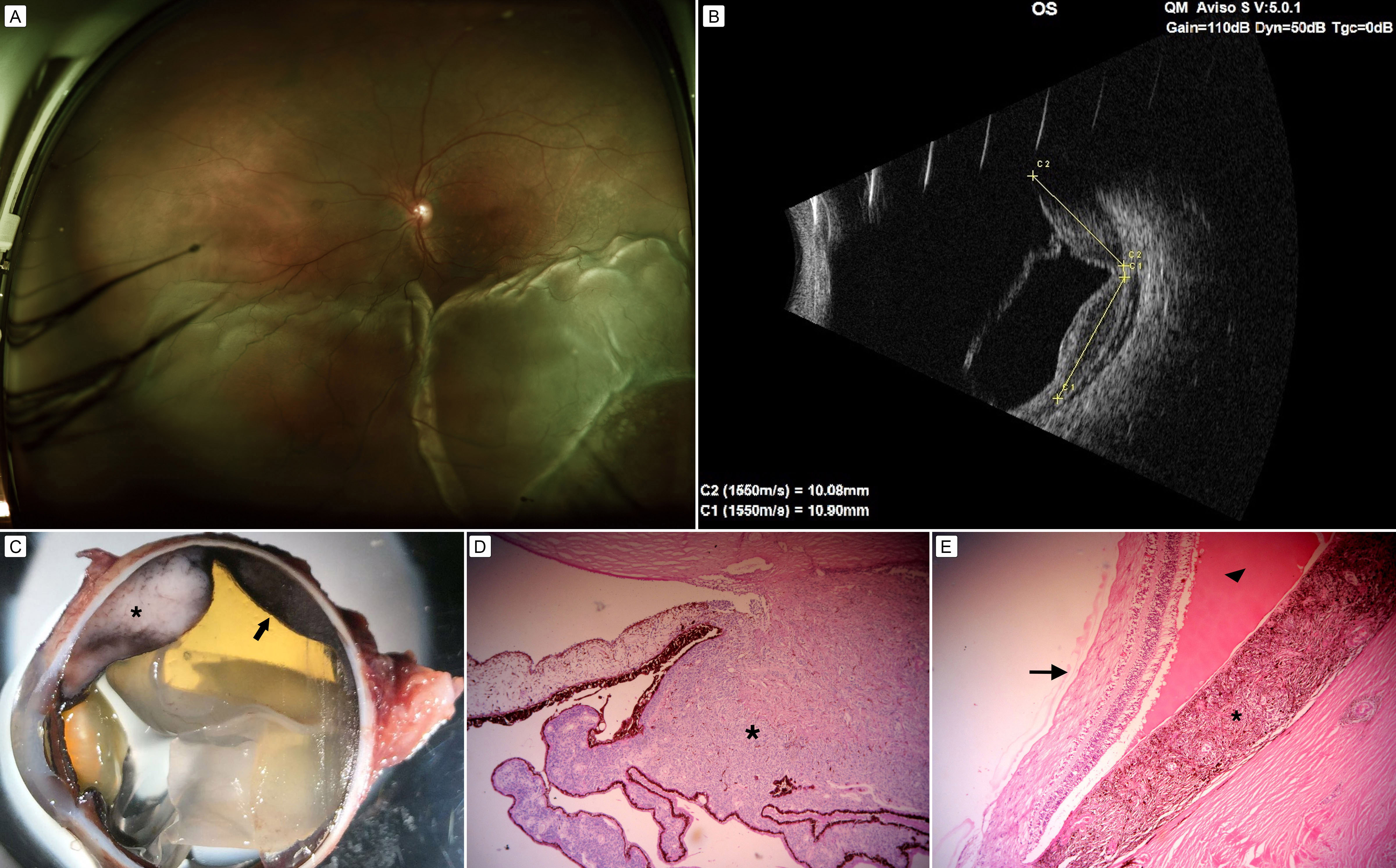
A 56-year-old man presented at the Asociación para Evitar la Ceguera en México with an inferior, exudative retinal detachment secondary to a uveal melanoma (A). Echography revealed a bilobulated choroidal solid tumor with medium-to-high reflectivity and retinal detachment (B). Enucleation was performed. The macroscopic appearance showed an amelanotic neoplasm in the ciliary body (asterisk) and a pigmented tumor (arrow) in the choroid with the overlying retina detached (C). Histopathologic examination (D,E [hematoxylin-eosin, original magnification ×200]) revealed an amelanotic tumor with widening of the ciliary body at the pars plicata (D, asterisk) and a pigmented neoplasm (E, asterisk) with the associated exudative (E, arrowhead) detachment of the retina (E, arrow).
Downloads
Article Details

This work is licensed under a Creative Commons Attribution-NonCommercial-NoDerivatives 4.0 International License.