Corneal dermoid
Article Sidebar
Main Article Content
Abstract
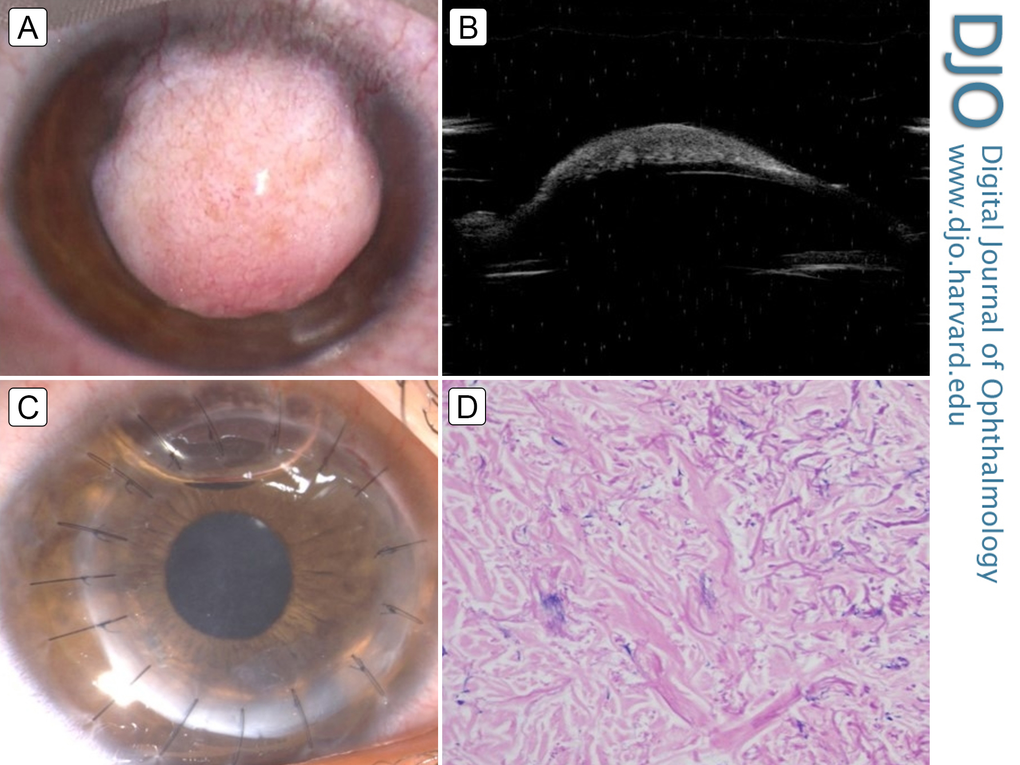
A 10-year-old boy presented at Advanced Eye Centre, Postgraduate Institute of Medical Education and Research, Chandigarh, for evaluation of a mass lesion in the right eye that had been gradually increasing in size since birth. Slit-lamp evaluation revealed a well circumscribed, lightly vascularized, elevated pinkish dermoid measuring 6.5 × 7.5 mm over the central cornea (A). No lid abnormalities, preauricular skin tags, or vertebral anomalies suggestive of Goldenhar syndrome were noted. On ultrasound biomicroscopy, the dermoid was seen to extend to the mid-stroma, with sparing of the deeper stroma and corneal endothelium (B). Deep anterior lamellar keratoplasty was performed, and postoperatively a clear corneal graft was achieved (C). Postoperative visual acuity was counting fingers at 1 meter, because of amblyopia, for which occlusion therapy of the normal left eye was initiated. Histopathological examination of the specimen revealed densely collagenized stroma with collagen bundles and nonkeratinized stratified squamous epithelium (D). Absence of significant fibroblastic invasion helped exclude corneal keloid, which usually occurs following trauma but which may rarely be congenital and simulate a corneal dermoid. Corneal dermoids, which are the least common of all ocular dermoid lesions (5%-6%), may be a rare cause of congenital corneal opacification and hence visual deprivation.
Downloads
Article Details

This work is licensed under a Creative Commons Attribution-NonCommercial-NoDerivatives 4.0 International License.