Cogan-Reese syndrome: image analysis with specular microscopy, optical coherence tomography, and ultrasound biomicroscopy
Article Sidebar
Main Article Content
Abstract
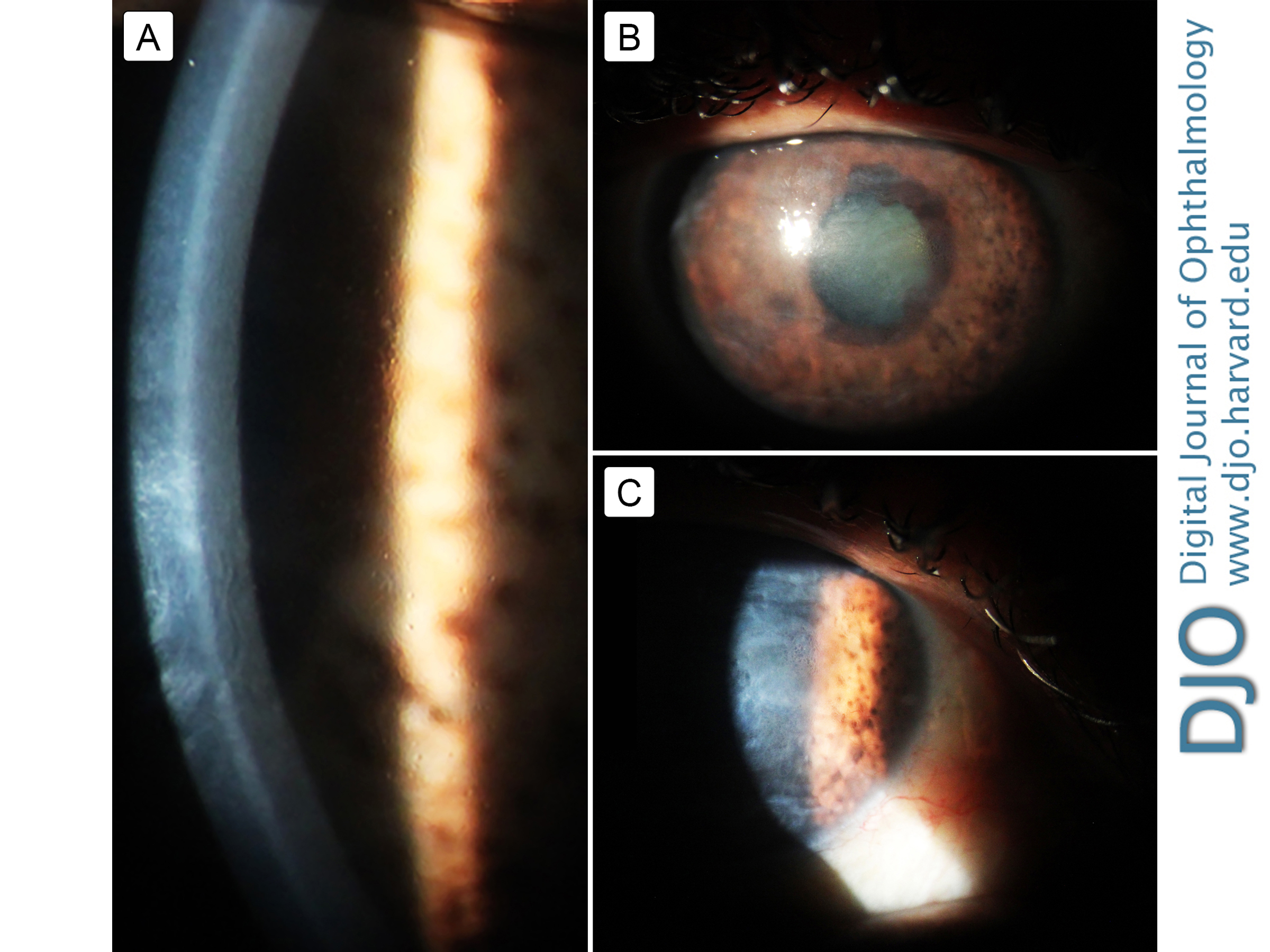
Iridocorneal endothelial (ICE) syndrome is a progressive clinical spectrum of corneal endothelial abnormalities affecting the cornea, iris, and iridocorneal angle. Three clinical variations are recognized: essential (progressive) iris atrophy, Chandler syndrome, and Cogan-Reese syndrome. Direct slit-lamp visualization of the cornea and anterior segment in cases of ICE syndrome is inadequate for precise and objective assessment of the affected structures. We describe the evolution of corneal and anterior segment structural changes in a woman with Cogan-Reese syndrome using three different methods of image analysis: specular microscopy, anterior segment optical coherence tomography, and ultrasound biomicroscopy.
Downloads
Article Details

This work is licensed under a Creative Commons Attribution-NonCommercial-NoDerivatives 4.0 International License.


