Lattice corneal dystrophy
Article Sidebar
Main Article Content
Abstract
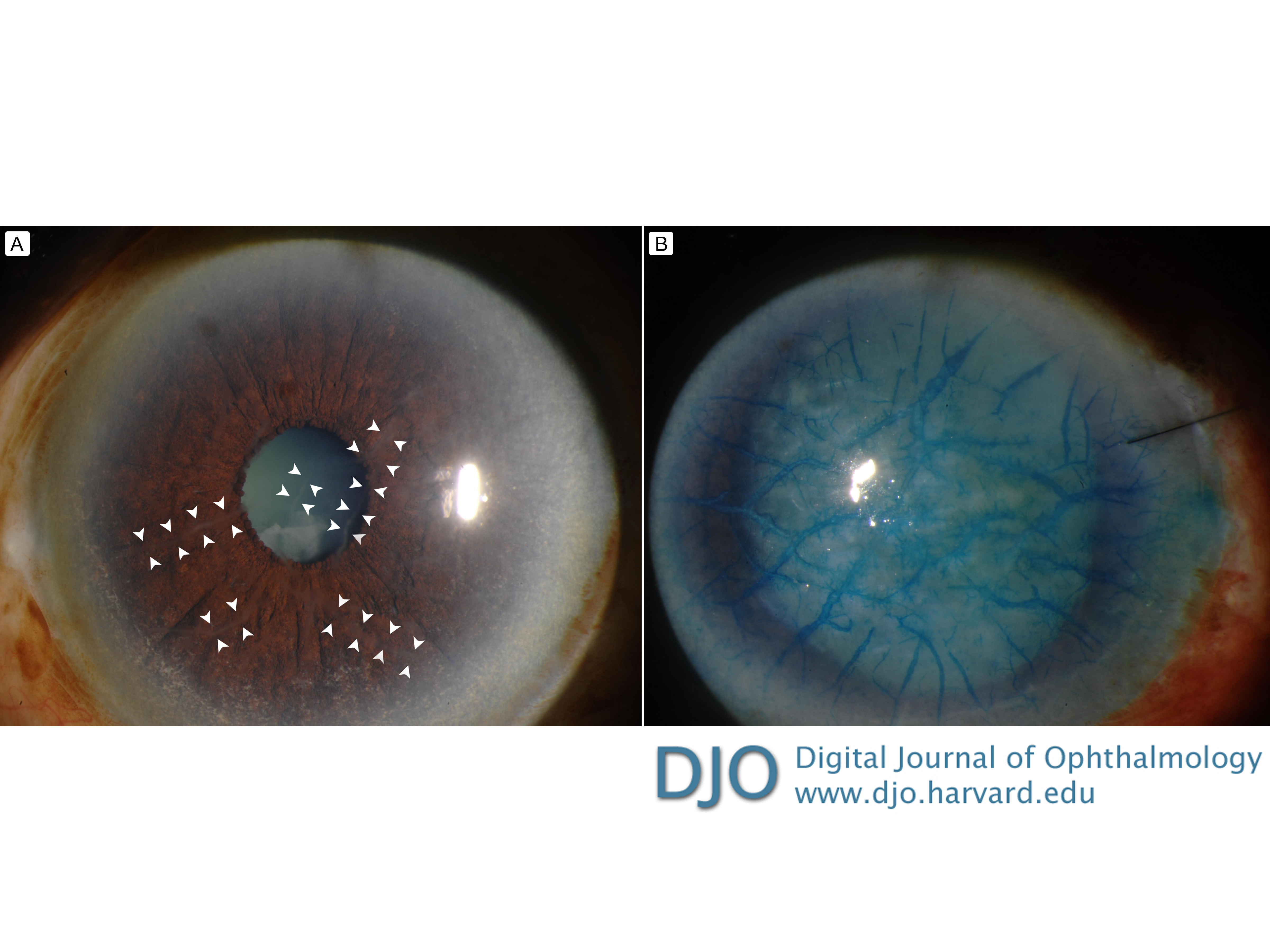
A 77-year-old woman with a history of glaucoma presented at the Wilmer Eye Institute with decreased vision of several months’ duration. She had also experienced occasional episodes of redness, itchiness, and burning sensation prior to presentation. On examination, the best-corrected visual acuity was 20/80 in the right eye and 20/25 in the left eye. Anterior segment examination of the right eye revealed a cataract and subtle opaque streaks in the corneal stroma (A, arrowheads). The remainder of the examination was unremarkable. Cataract surgery was planned, but was complicated by severe intraocular pressure elevation refractory to anterior chamber taps and intravenous mannitol, resulting in corneal haziness. To help visualize the lens capsule intraoperatively, trypan blue was injected into the anterior chamber of the eye. With significant and persistent obscuration of the surgical view, the surgery was aborted. Postoperative examination showed peculiar staining of the radially oriented, branching, refractile, filamentous, subepithelial and stromal opacities by trypan blue (B).
Downloads
Article Details

This work is licensed under a Creative Commons Attribution-NonCommercial-NoDerivatives 4.0 International License.