Presumed topiramate-induced retinopathy in a 58-year-old woman
Article Sidebar
Main Article Content
Abstract
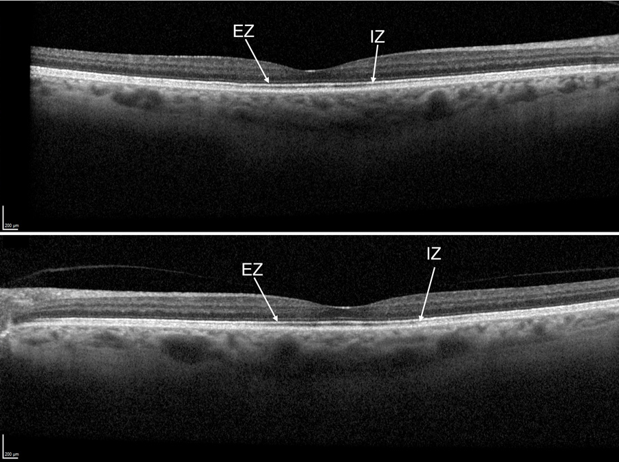
We present a case of presumed topiramate-induced retinopathy in a 58-year-old woman who presented with progressive, bilateral visual loss following a 3- to 4-year history of oral topiramate intake for migraine. She reported difficulty with light adaptation, hemeralopia, and color desaturation. Her best-corrected visual acuity was 1/60 (20/1200) in the right eye and 6/18 (20/60) in the left eye, and she performed poorly on Ishihara color plate testing. Anterior segment examination was normal; dilated funduscopy showed mild macular pigmentary changes. Optical coherence tomography revealed subtle thinning and reduced reflectivity of the subfoveal ellipsoid zone and interdigitation zone bilaterally, associated with increased foveal autofluorescence. Humphrey visual field 24-2 revealed central defects. Electrodiagnostic testing showed a reduced and delayed b-wave and a normal a-wave on photopic full-field electroretinogram (ERG), with normal scotopic responses; multifocal ERG revealed reduced responses in the inner 10° in both eyes. She underwent extensive investigations including whole-body computed tomography and positron emission tomography scan, magnetic resonance imaging of the brain, uveitis screening, retinal autoantibody testing, and genetic testing on the retinal dystrophy panel to rule-out other causes for her presentation, all of which were normal or negative.
Downloads
Article Details

This work is licensed under a Creative Commons Attribution-NonCommercial-NoDerivatives 4.0 International License.


References
Minton GC, Miller AD, Bookstaver PB, Love BL. Topiramate: safety and efficacy of its use in the prevention and treatment of migraine. J Cent Nerv Syst Dis 2011;3:155-68. DOI: https://doi.org/10.4137/JCNSD.S4365
Brown SD, Wolf HH, Swinyard EA, et al. The novel anticonvulsant topiramate enhances GABA-mediated chloride flux. Epilepsia 1993;34:S122-3.
White HS, Brown SD, Woodhead JH, et al. Topiramate enhances GABA-mediated chloride flux and GABA-evoked chloride currents in murine brain neurons and increases seizure threshold. Epilepsy Res 1997;28:167-79. DOI: https://doi.org/10.1016/S0920-1211(97)00045-4
Gibbs JW 3rd, Sombati S, DeLorenzo RJ, Coulter DA. Cellular actions of topiramate: blockade of kainate-evoked inward currents in cultured hippocampal neurons. Epilepsia 2000;41(S1):10-6. DOI: https://doi.org/10.1111/j.1528-1157.2000.tb02164.x
DeLorenzo RJ, Sombati S, Coulter DA. Effects of topiramate on sustained repetitive firing and spontaneous recurrent seizure discharges in cultured hippocampal neurons. Epilepsia 2000;41:S40-44. DOI: https://doi.org/10.1111/j.1528-1157.2000.tb06048.x
Richa S, Yazbek JC. Ocular adverse effects of common psychotropic agents. CNS Drugs 2010;24:501-26. DOI: https://doi.org/10.2165/11533180-000000000-00000
Cyrlin MN. Primary and secondary angle-closure glaucomas. In: Samples J, Schacknow P, eds. Clinical Glaucoma Care. New York, NY: Springer; 2014:287-322. DOI: https://doi.org/10.1007/978-1-4614-4172-4_14
Roff Hilton EJ, Hosking SL, Betts T. The effect of antiepileptic drugs on visual performance. Seizure 2004;13:113-28. DOI: https://doi.org/10.1016/S1059-1311(03)00082-7
Yeung TLM, Li PSH, Li KKW. Presumed topiramate retinopathy: a case report. J Med Case Rep 2016;10:1-4. DOI: https://doi.org/10.1186/s13256-016-0980-x
Vaphiades MS, Mason J. Foggy visual field defect. Surv Ophthalmol 2004;49:266-7. DOI: https://doi.org/10.1016/j.survophthal.2003.12.004
Asensio-Sánchez VM, Torreblanca-Agüera B, Martínez-Calvo S, Calvo MJ, Rodríguez R. Severe ocular side effects with Topamax [in Spanish]. Arch Soc Esp Oftalmol 2006;81:345-8. DOI: https://doi.org/10.4321/S0365-66912006000600010
Beyenburg S, Weyland C, Reuber M. Presumed topiramate-induced maculopathy. Epilepsy Behav 2009;14:556-9. DOI: https://doi.org/10.1016/j.yebeh.2008.12.015
Severn PS, Symes R, Rajendram R, Pal B. Topiramate maculopathy secondary to dose titration: first reported case. Eye 2015;29:982. DOI: https://doi.org/10.1038/eye.2015.45
Gualtieri W, Janula J. Topiramate maculopathy. Int Ophthalmol 2013;33:103-6. DOI: https://doi.org/10.1007/s10792-012-9640-3
Tsui I, Casper D, Chou CL, Tsang SH. Electronegative electroretinogram associated with topiramate toxicity and vitelliform maculopathy. Doc Ophthalmol 2008;116:57-60. DOI: https://doi.org/10.1007/s10633-007-9084-7
Johnson AA, Guziewicz KE, Lee CJ, et al. Bestrophin 1 and retinal disease. Prog Retin Eye Res 2017;58:45-69. DOI: https://doi.org/10.1016/j.preteyeres.2017.01.006
Hadjikoutis S, Morgan JE, Wild JM, Smith PEM. Ocular complications of neurological therapy. Eur J Neurol 2005;12:499-507. DOI: https://doi.org/10.1111/j.1468-1331.2005.01025.x
Krauss G. L, Johnson M. A, Sheth S, Miller N. R. A controlled study comparing visual function in patients treated with vigabatrin and tiagabine. Journal of neurology, neurosurgery, and psychiatry 2003;74, 339–343. DOI: https://doi.org/10.1136/jnnp.74.3.339
Miller NR, Johnson MA, Paul SR, et al. Visual dysfunction in patients receiving vigabatrin: clinical and electrophysiologic findings. Neurology 1999;53:2082-7. DOI: https://doi.org/10.1212/WNL.53.9.2082
Johnson MA, Krauss GL, Miller NR, et al. Visual function loss from vigabatrin. Neurology 2000;55:40-5. DOI: https://doi.org/10.1212/WNL.55.1.40
Kjellström S, Bruun A, Isaksson B, et al. Retinal function and histopathology in rabbits treated with topiramate. Doc Ophthalmol 2006;113:179-86. DOI: https://doi.org/10.1007/s10633-006-9027-8
Kapousta-Bruneau NV. Opposite effects of GABA(A) and GABA(C) receptor antagonists on the b-wave of ERG recorded from the isolated rat retina. Vision Res 2000;40:1653-65. DOI: https://doi.org/10.1016/S0042-6989(00)00028-6
Zhu M, Krilis M, Gillies MC. The relationship between inner retinal cavitation, photoreceptor disruption, and the integrity of the outer limiting membrane in macular telangiectasia type 2. Retina 2013;33:1547-50. DOI: https://doi.org/10.1097/IAE.0b013e318285cb9c