Retinal detachment in combined hamartoma of the retina and retinal pigment epithelium
Article Sidebar
Main Article Content
Abstract
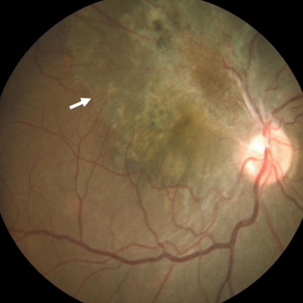
A 30-year-old man presented at Sankara Nethralaya with a complaint of diminished vision in the right eye of 2 years’ duration. On examination, his best-corrected visual acuity was 20/400. Dilated fundus examination revealed an ill-defined, grayish lesion, with hyperpigmentation and retinal vascular tortuosity. Retinal folds from the inferotemporal aspect of the lesion extended outward radially, blending with the surrounding tissue but clearly delimited (A [arrow] and B [red-free photograph]). Optical coherence tomography (OCT) of the right eye through the lesion showed partial-thickness disorganization of the retinal layers (C). OCT through the macula showed epiretinal membrane and retinal detachment (D). The patient was diagnosed with retinal detachment and combined hamartomas of the retina and retinal pigment epithelium.
Legends
Figure:1a) Fundus photo of the right eye showing CHR-RPE with epiretinal membrane and retinal detachment with demarcation line (white arrow), b) Red free photograph for better visualisation c) OCT through the lesion showing partial-thickness disorganisation of retinal layers, d) OCT through the macula and disc showing epiretinal membrane (asterisk), obscuration of the normal retinal layers and retinal detachment.
Downloads
Article Details

This work is licensed under a Creative Commons Attribution-NonCommercial-NoDerivatives 4.0 International License.